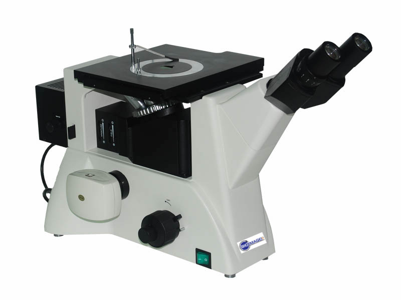
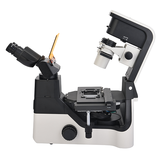
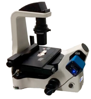
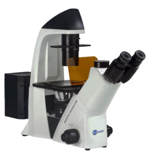
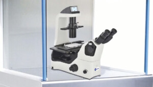
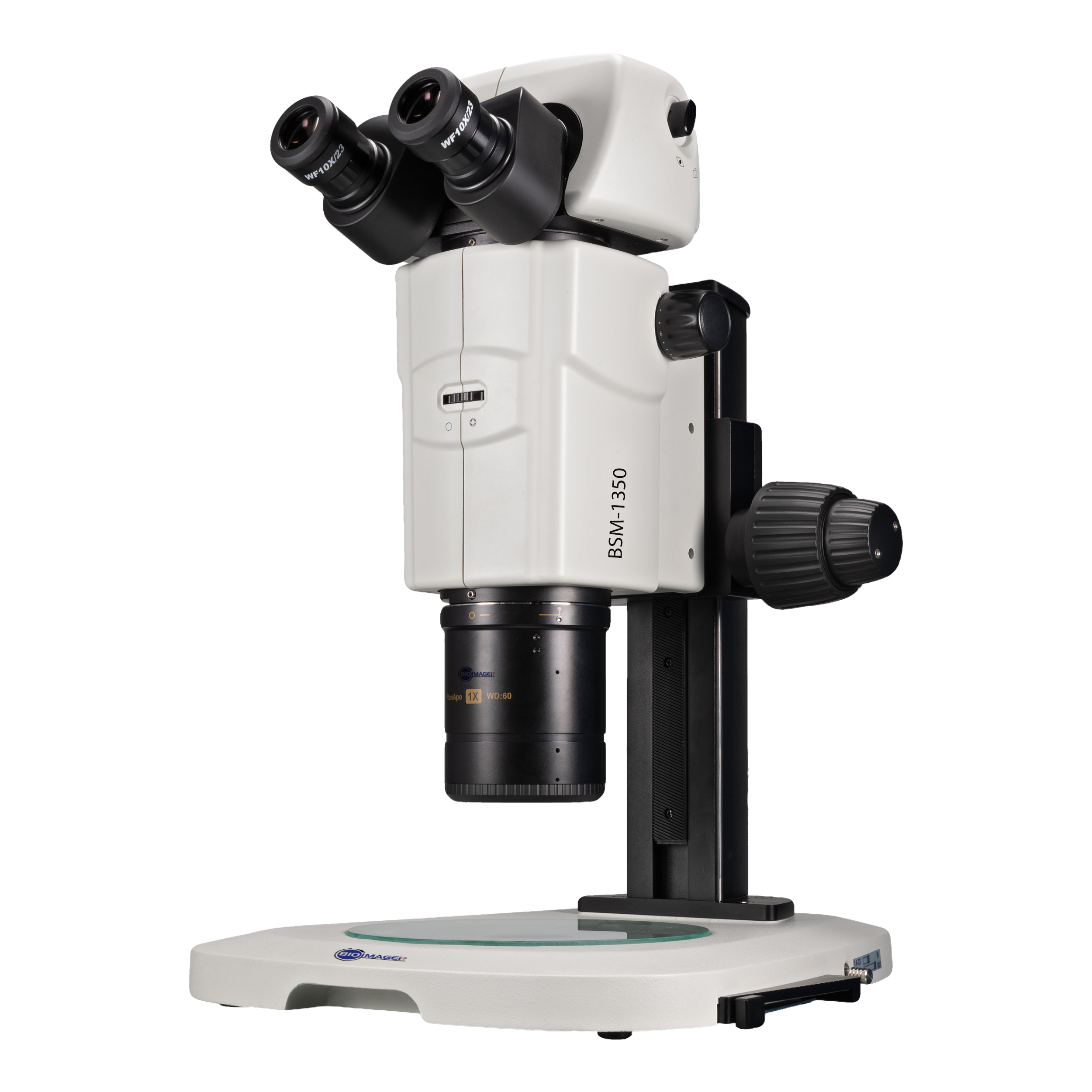
BIOIMAGER's inverted fluorescence microscopes are available at low price, versatile designs and configurations based on your requirements and budget.Continue Reading...
Discover a New Dimension of Imaging: Unleash the Power of Inverted Fluorescence Microscopy
If you are dealing with tissue cell culture, handling liquids in your device, or working with a microfluidics chamber, an inverted biological microscope is an essential tool to have. One likes to make a video of living cells to know how they grow, divide or die and the rate, you need an inverted fluorescence microscope, a digital camera and software for the time-lapse imaging.
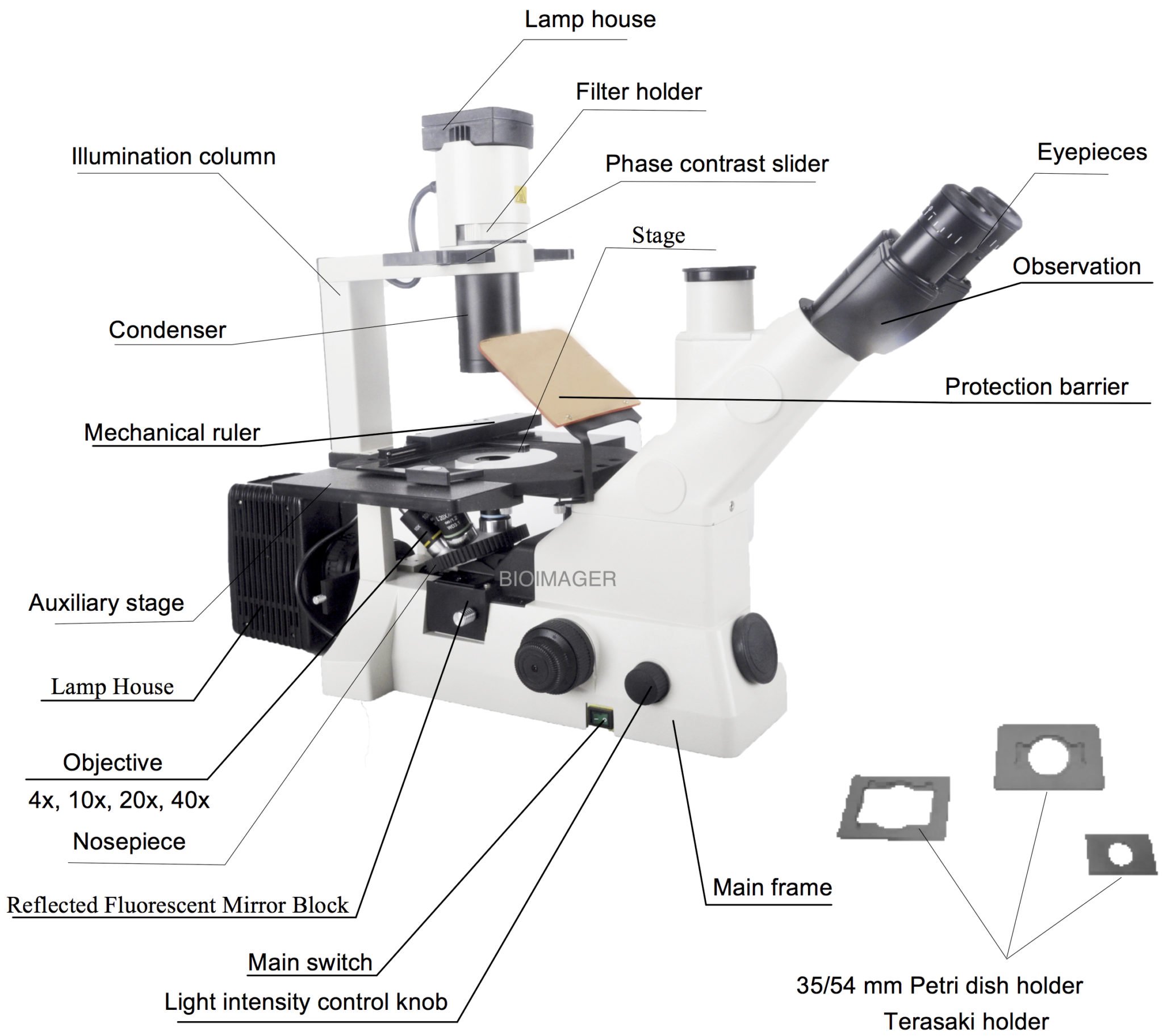
An inverted fluorescence microscope is a type of microscope that is commonly used in biological research to observe living cells or tissues. In contrast to a traditional upright microscope, the inverted microscope has its objective lens facing downwards, while the specimen is mounted on a stage that is positioned above the objective. This arrangement allows for the observation of thicker samples, as well as the ability to manipulate the specimen from above.
Inverted fluorescence microscopes are equipped with specialized optics and light sources that allow for the visualization of fluorescently labelled specimens. This type of microscopy is widely used in cell biology and genetics research to observe and manipulate fluorescently labelled proteins, DNA, and other cellular structures. In addition to live cell imaging, inverted fluorescence microscopes are also used for fixed specimen imaging, such as in immunofluorescence experiments.
Overall, the inverted fluorescence microscope is a powerful tool for the visualization and analysis of biological samples, offering high-resolution imaging of living and fixed specimens.
What to consider in selecting an inverted fluorescence microscope?
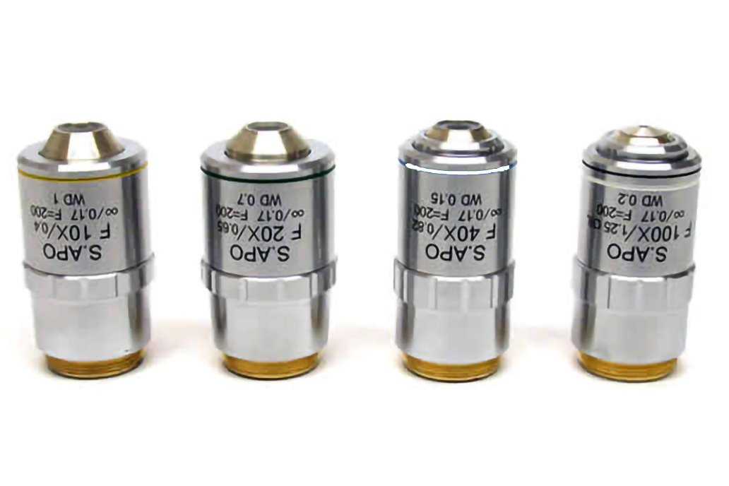
Objective lenses-magnification:
normally the inverted microscopes come with 10x, 20x and 40x obj lenses, some models include 4x. A 4x obj lens allows seeing an entire field of a well in a 96-well plate. You can get objective lenses from 1.25x till 150x and even more.
Objective lenses and their working distance:
Working distance (WD) of an objective lens is defined as a distance from the edge of the lens until the specimen. If you use a 100x/NA 1.25/WD: 0.12mm to see tissue culture cells inside a well of a 96-well plate, you will see nothing! Why? Because the thickness of well plate plastic is around 0.5mm which is longer than lens WD=0.12mm.
Phase Contrast:
generally speaking 10x, 20x and 40x obj lenses with Phase Contrast are available. Depending on the model you can expect a 4x Ph as well.
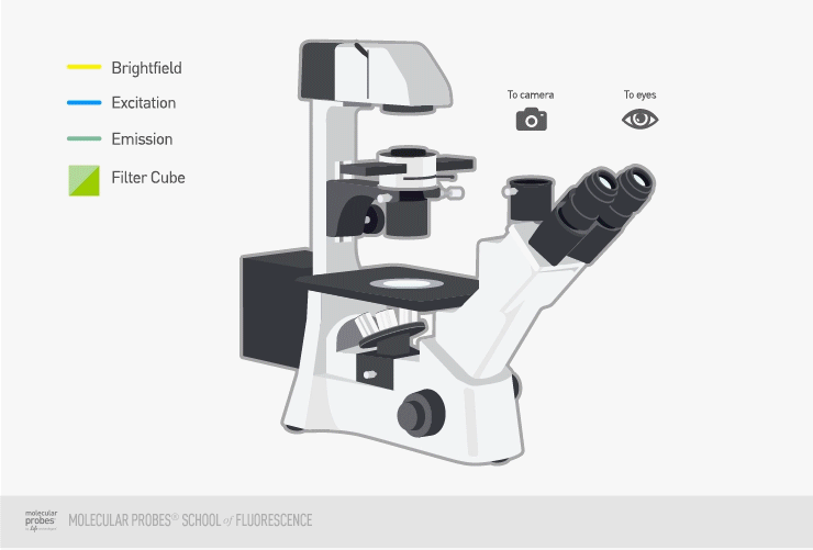
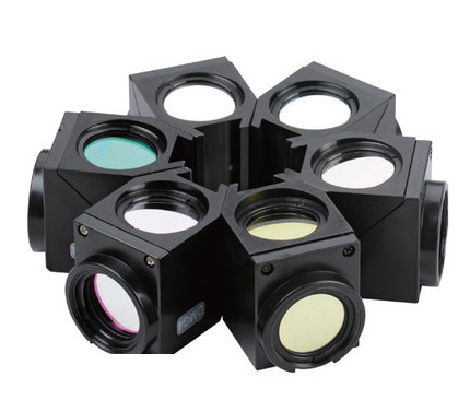
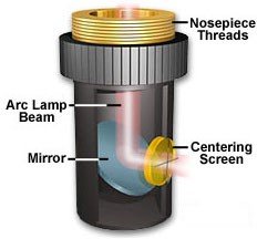
Filter slider vs. Filter wheel:
Both filter sliders and filter wheels are used in fluorescence microscopes to select and switch between different filters that are required for specific fluorescence imaging applications.
A filter slider is a mechanism that contains several filters that can be moved back and forth in front of the light source or the detector. The user manually selects the required filter by sliding the filter slider into position. Filter sliders are simple and easy to use, but they can be slow and may cause vibrations and misalignment in the microscope optics.
A filter wheel, on the other hand, is a circular device that contains multiple filters arranged in a circular pattern. The filter wheel rotates to position the desired filter in front of the light source or the detector. Filter wheels can be automated and controlled by computer software, allowing for faster and more precise filter selection. They also eliminate vibrations and misalignment caused by filter sliders.
In general, filter wheels are more advanced and preferred over filter sliders in modern fluorescence microscopes because of their speed, precision, and reliability. However, filter sliders are still used in some applications where the number of filters required is small, and the cost of a filter wheel system may not be justified.
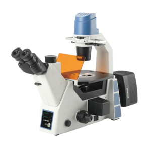
Among our inverted fluorescence microscope, BIM800FLW has a filter wheel. We have a newer technology which uses neither filer slider nor filter wheel. That uses indeed a multi-band filter set in which the light source is selected digitally. Check our IncuScope microscopes, particularly IS25-BG model.
LED or HBO:
LED and HBO (high-pressure mercury) lamps are two common light sources used in fluorescence microscopes. Each has its advantages and disadvantages, and the choice between them depends on the specific requirements of the experiment.
LEDs have become increasingly popular as a light source for fluorescence microscopes due to their versatility, low power consumption, long lifespan, and low heat output. They can be easily switched on and off, and their output can be precisely controlled. LED illumination is also ideal for multicolor imaging, as multiple LEDs with different wavelengths can be used to excite different fluorophores. LED light sources are typically less expensive than HBO lamps, and they don’t require frequent bulb replacements.
HBO lamps, on the other hand, have been used for decades in fluorescence microscopy and provide intense, stable, and broadband illumination across the UV to red spectral range. They can be used to excite a wide range of fluorophores, and they are particularly useful for high-resolution imaging and time-lapse experiments. However, they require a warm-up period, and their bulb has a limited lifespan and requires periodic replacement. HBO lamps are also more expensive than LED light sources, and they generate more heat, which can affect sample viability and stability.
In summary, both LED and HBO lamps have their advantages and disadvantages, and the choice between them depends on the specific experimental requirements and budget constraints. LED light sources are generally preferred for their versatility, low heat output, and low cost, while HBO lamps are preferred for high-resolution imaging and long-term time-lapse experiments.
Upgrade:
There are several ways to upgrade a fluorescence microscope to improve its performance, functionality, and versatility. Some common upgrades include:
Upgrading the light source: Replacing the current light source, such as a mercury lamp, with a more advanced and efficient one, such as an LED light source, can improve illumination quality and reduce heat generation.
Adding a filter wheel or filter slider: Installing a filter wheel or slider system allows for fast and precise selection of the desired filters, enabling multicolor imaging and reducing alignment errors.
Adding a motorized stage: A motorized stage allows for automated and precise movement of the sample, enabling high-throughput imaging and 3D imaging.
Adding a camera: Upgrading the camera system, such as replacing the CCD camera with a more sensitive sCMOS camera, can improve the signal-to-noise ratio and increase image resolution and speed.
Adding advanced imaging techniques: Adding advanced imaging techniques, such as confocal microscopy, super-resolution microscopy, or fluorescence lifetime imaging microscopy (FLIM), can provide additional information about the sample and improve imaging resolution and accuracy.
Upgrading the software: Upgrading the microscope software, such as adding new features or improving image processing algorithms, can enhance data analysis and visualization capabilities.
These upgrades can be expensive, and the cost depends on the specific upgrade and the microscope model. However, they can significantly improve the microscope’s performance and expand its capabilities, enabling researchers to address more complex and challenging biological questions.
Think of long future use of the microscope. Can you upgrade it to have more features? Having more filters, switching from HBO to LED, more spots for extra obj lenses, might be having polarized, darkfield, and probably DIC Nomarski are examples of the questions we ask our clients before suggesting a model.
BIOIMAGER’s inverted fluorescence microscopes are available at low price and versatile designs and configurations. It comes with trinocular head or side video port to attach a CCD digital camera, to have a live view of your cells or any stained fluorescent specimen. We offer over 40 filters from UV to far red with HBO, LED or Xenon illumination.
Customized filters are available for all epi-fluorescence microscope models.Contact us for details.
Please select the products based on your requirements from the left side list or below table:
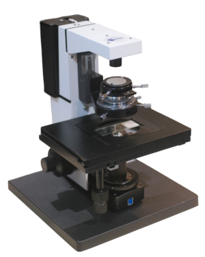
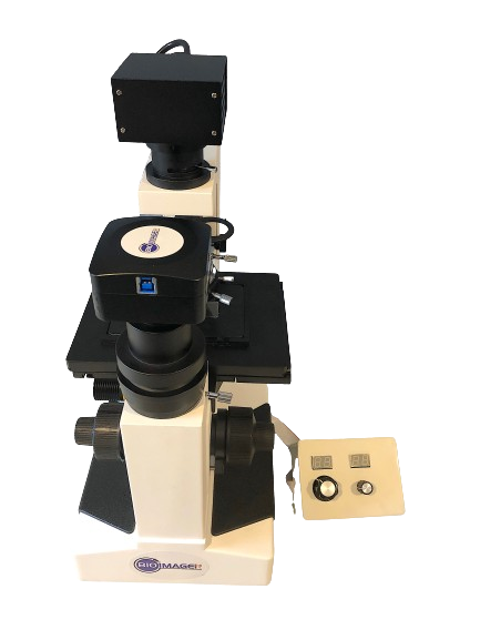
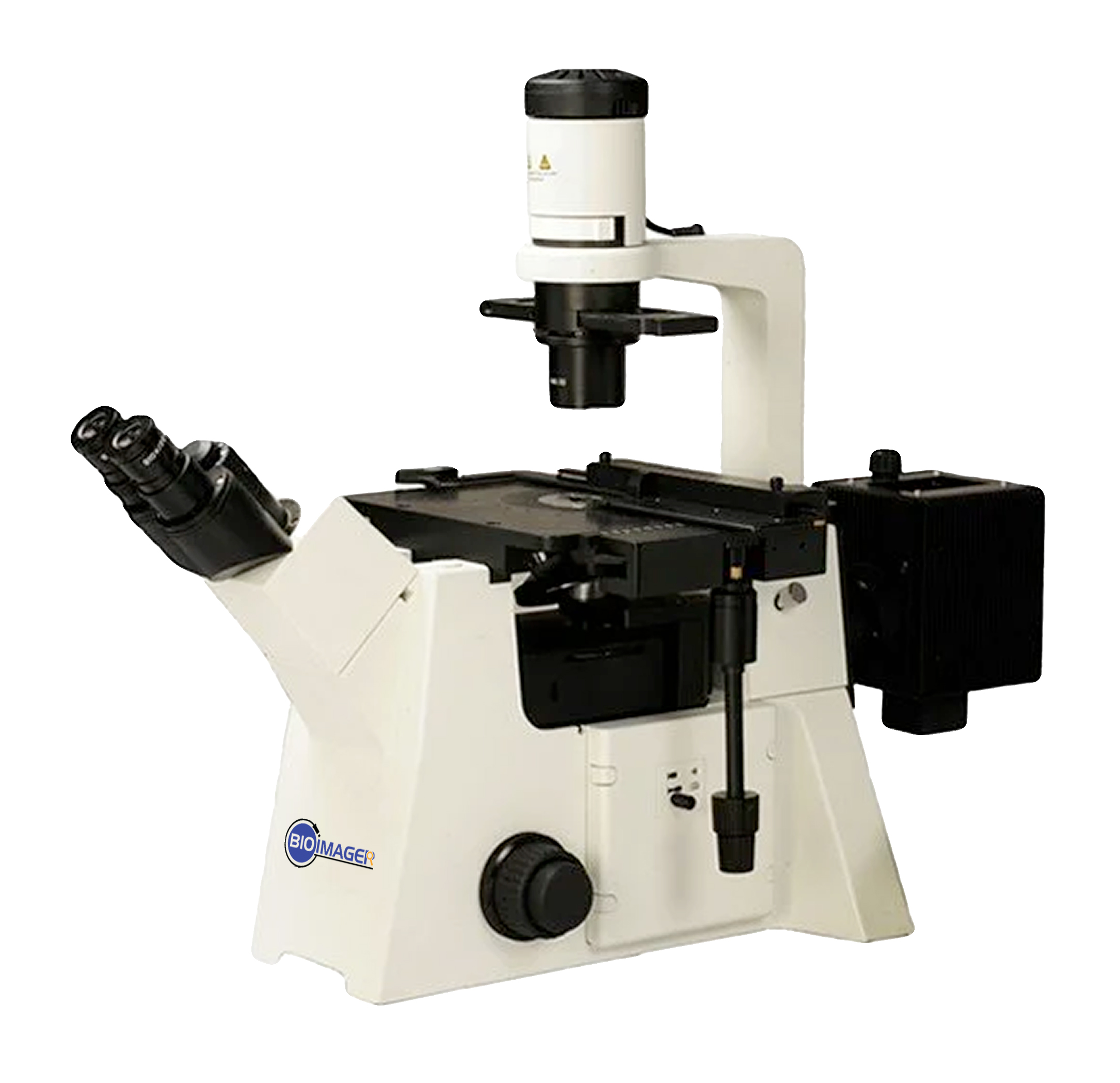
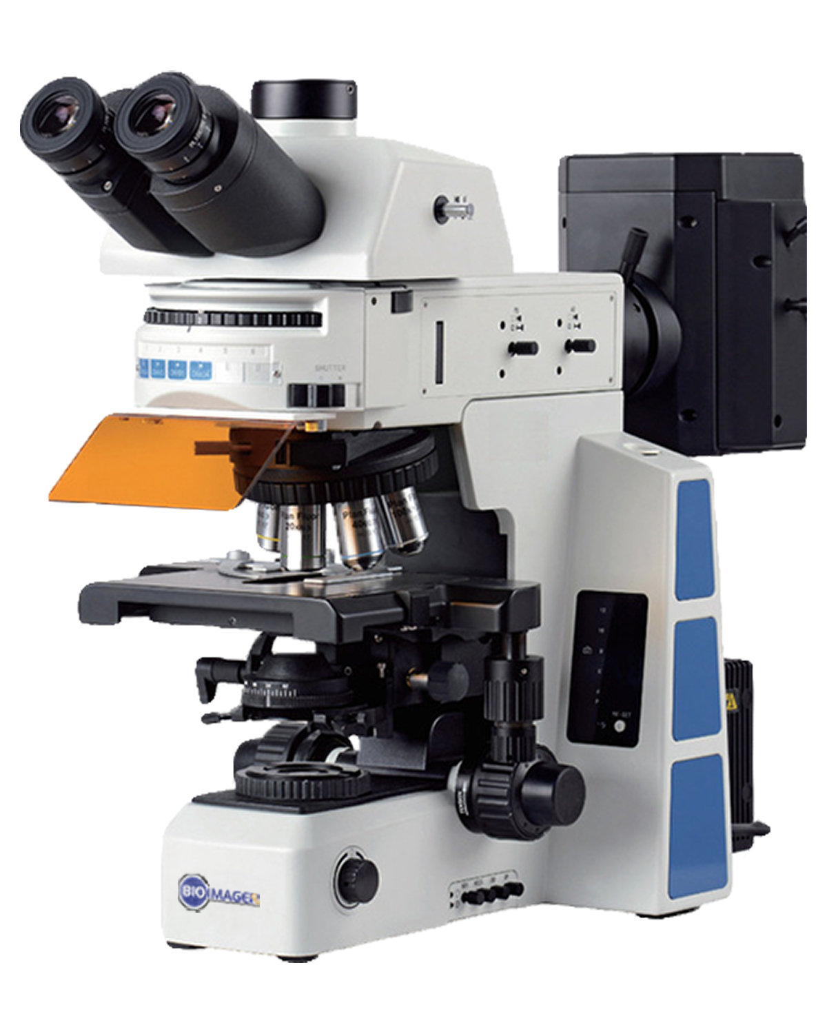
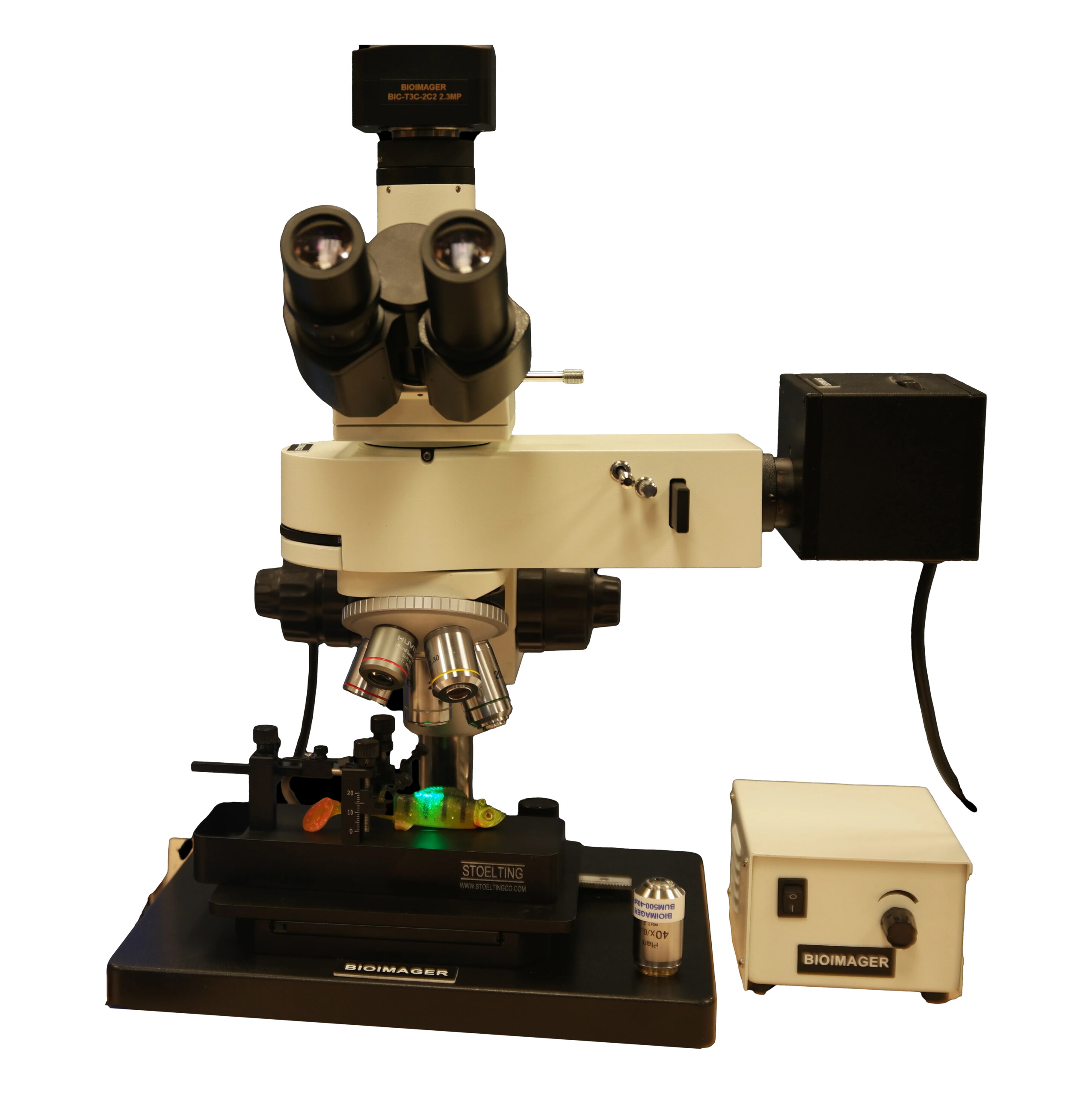
Details of a representative inverted fluorescence microscope:
Applications:
Inverted fluorescence microscopy has a wide range of applications in life sciences, particularly in cell biology and microbiology. Some common applications include:
Live cell imaging: Inverted fluorescence microscopy allows for the observation of living cells in real-time, enabling researchers to study cell behavior and interactions in a non-invasive manner.
Fluorescence labeling: The technique is commonly used to visualize and study fluorescently labeled molecules such as proteins, DNA, and RNA in cells and tissues.
Immunofluorescence: Inverted fluorescence microscopy can be used to visualize the distribution and expression of specific proteins in cells and tissues using immunofluorescence labeling.
Fluorescence resonance energy transfer (FRET): The technique can be used to study molecular interactions and protein-protein interactions by monitoring FRET between two fluorophores.
Time-lapse imaging: Inverted fluorescence microscopy allows for the acquisition of time-lapse images, which can be used to study dynamic processes such as cell division, migration, and differentiation.
Microbial imaging: The technique can be used to study microbial communities, including biofilms, by visualizing fluorescently labeled microbes.
Overall, inverted fluorescence microscopy is a versatile technique that can be used to study a wide range of biological processes and applications, enabling researchers to gain a deeper understanding of life at the cellular and molecular levels.
Examples of our customers:
Canadian Blood Services, Ottawa, Ontario
Canada
Concordia University, Toronto, Ontario
Héma-Québec, Québec, QC
Sick Kids Hospital, Toronto, Ontario
University of British Columbia (UBC), Vancouver, BC