Cell Culture
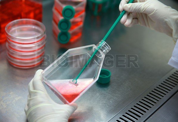
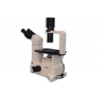
Suitable microscopes for tissue cell cultures are inverted biological fluorescent microscopes with phase contrast, DIC, fluorescnce imaging in time-lapse modeContinue Reading...
Showing all 8 results
-
-
-
$USD 9,750.00 Add to cart
-
-
$USD 8,875.00 Add to cart
-
-
$USD 4,395.00 Add to cart
-
Showing all 8 results
What microscope is good for tissue cell culture imaging?
When it comes to imaging cell cultures, several types of microscopes can be used depending on the specific requirements of the experiment or analysis. Here are a few common microscope options for cell culture imaging:
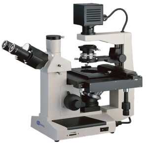
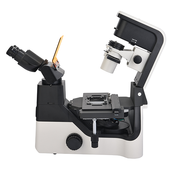
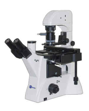
- Inverted Microscope: Inverted microscopes are commonly used for cell culture imaging. They have a unique design where the objective lens is positioned beneath the specimen stage, allowing you to observe cells from the bottom of the culture dish or flask. Inverted microscopes are ideal for long-term live cell imaging, as they provide stability and accessibility for manipulating the culture while maintaining optimal environmental conditions.
- Fluorescence Microscope: Fluorescence microscopy is widely used in cell biology research. Fluorescence microscopes are equipped with a specialized light source and filters that enable the visualization of specific fluorescent dyes or proteins within cells. This technique is particularly useful for studying cellular processes, protein localization, and cell signaling. In cell culture imaging, fluorescence microscopes are commonly used to observe specific cellular components, such as nuclei, cytoskeletal structures, or labeled proteins.
- Confocal Microscope: Confocal microscopy is an advanced imaging technique that provides high-resolution, three-dimensional images of cells and tissues. It uses a pinhole to eliminate out-of-focus light, resulting in improved contrast and optical sectioning capabilities. Confocal microscopes are particularly useful for imaging thick cell cultures or visualizing cellular dynamics over time. They can also generate detailed 3D reconstructions of cellular structures.
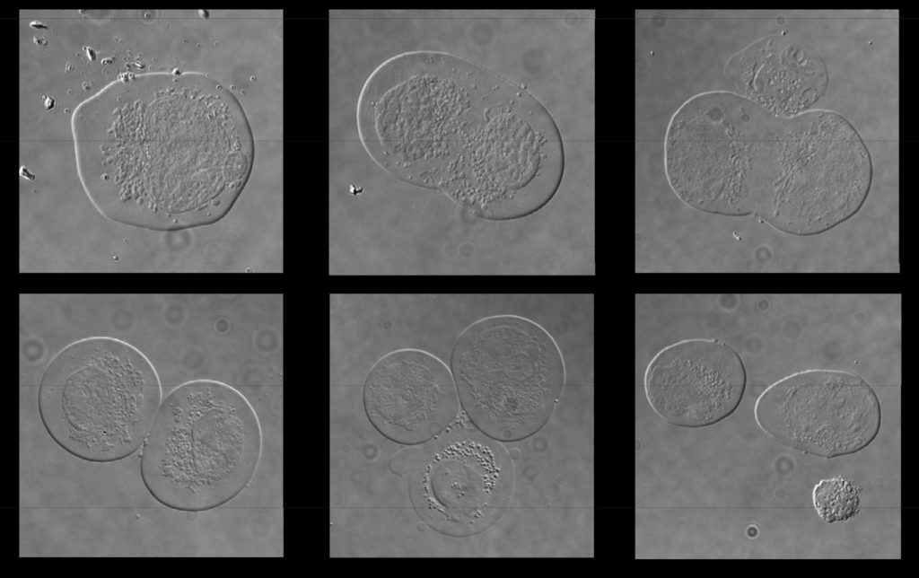
- Phase-Contrast Microscope: Phase-contrast microscopy is a technique that enhances the contrast of transparent specimens, such as live cells, without the need for staining or labeling. It converts subtle changes in the refractive index of the specimen into variations in brightness, allowing you to visualize cellular structures and movements. Phase-contrast microscopes are commonly used in routine cell culture monitoring and observation.
- Differential Interference Contrast (DIC) Microscope: DIC microscopy is another contrast-enhancing technique that produces detailed, three-dimensional images of transparent specimens. It uses polarized light to create a relief-like appearance, highlighting fine structural details in cells. DIC microscopes are particularly useful for visualizing cell morphology, organelles, and membrane structures.
- Time-Lapse Microscope: Time-lapse microscopes are specialized systems designed for capturing images or videos of cells over extended periods. These microscopes are often equipped with environmental control chambers to maintain optimal conditions for cell growth and imaging. Time-lapse microscopy allows for the observation of cellular dynamics, cell division, migration, and other time-dependent processes.
When choosing a microscope for cell culture imaging, consider factors such as resolution, magnification, imaging speed, compatibility with your experimental requirements, and budget constraints. Additionally, consult with an imaging experts to determine the most suitable microscope for your specific applications.