Microfluidics & MEMS
Showing all 5 results
-
$USD 120.00 Add to cart
-
-
 Request a Quote Request a Quote
 -
-
$USD 9,750.00 Add to cart
Showing all 5 results
Microscopes for Microfluidics and MEMS
When it comes to imaging microfluidics systems that incorporate microelectromechanical systems (MEMS), there are specific microscope options that are suitable for visualizing and analyzing these devices. Here are some microscope types commonly used for imaging microfluidics MEMS:
- Optical Microscope: An optical microscope is a versatile tool commonly used for imaging microfluidic MEMS devices. It allows for the visualization of device structures, fluid flow patterns, and the behavior of particles or cells within the microchannels. Optical microscopes can provide high-resolution imaging and can be equipped with various imaging techniques such as brightfield, darkfield, phase contrast, or fluorescence microscopy.
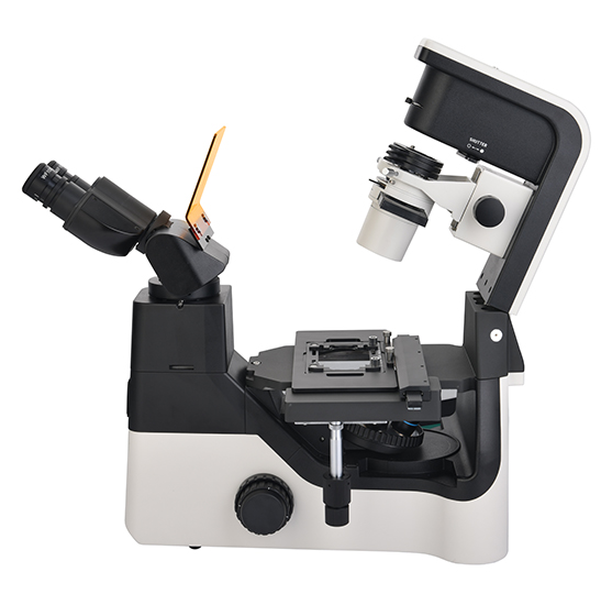
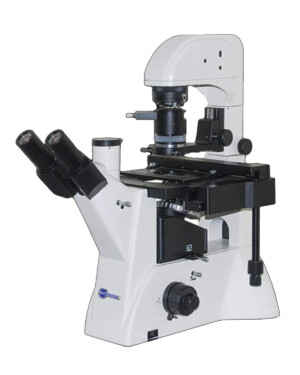
A. Inverted Microscopes
An inverted microscope is a commonly used tool for imaging microfluidic systems. It is particularly well-suited for observing and analyzing microfluidic devices because of its unique design, which allows for easy access to the fluidic channels and the ability to work with cell cultures or samples in a controlled environment.
| 1. Sample Compatibility: Inverted microscopes can accommodate a wide range of microfluidic devices, including glass or polymer-based chips, PDMS (polydimethylsiloxane) devices, or other custom-made microfluidic setups. The microscope stage typically features mechanical stability and compatibility with various sample holders or adapters for secure positioning of microfluidic chips. | 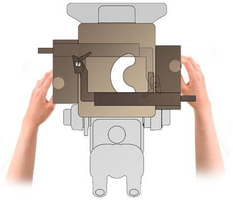 |
| 2. Fluorescence Imaging: Many inverted microscopes are equipped with fluorescence capabilities, which are highly valuable for microfluidics research. Fluorescent dyes, markers, or fluorescently tagged molecules can be employed within the microfluidic device, and the inverted microscope allows for the visualization and analysis of fluorescence signals. This facilitates the investigation of cellular processes, molecular interactions, or specific markers within microfluidic environments. | 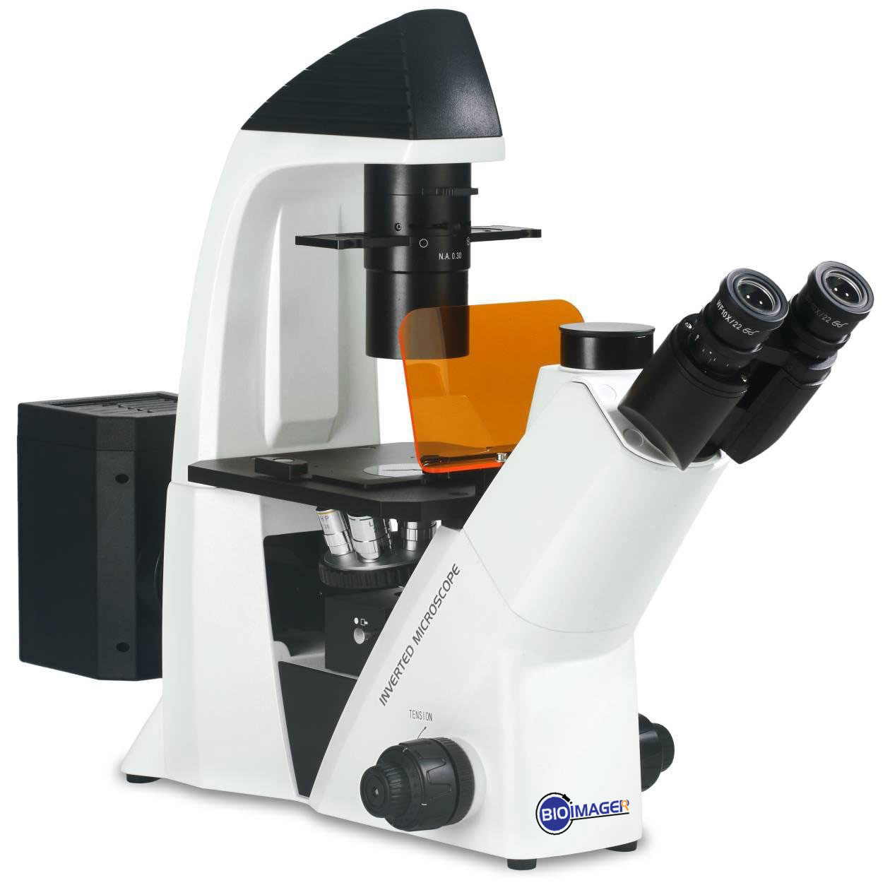 |
| 3. Time-Lapse Imaging: Inverted microscopes with motorized stages and environmental control chambers enable time-lapse imaging of microfluidic systems. This functionality allows for the capture of dynamic processes, such as fluid flow, droplet formation, particle manipulation, or cell migration, over extended time periods. High-speed imaging capabilities can also be incorporated to capture rapid events within microfluidic channels. |  |
B. Upright Microscopes
While inverted microscopes are commonly used for imaging microfluidic devices, upright microscopes can also be utilized in certain cases. Although not as common as inverted microscopes for microfluidics, upright microscopes offer unique advantages in specific experimental setups. Here are some considerations when using an upright microscope for microfluidic devices:
- Imaging Configuration: In an upright microscope, the objective lens is positioned above the sample stage, making it suitable for imaging from the top of microfluidic devices. This configuration is advantageous when the focus of the experiment is on the upper surface of the microfluidic channels or when using transparent substrates that allow direct imaging from above.
- Transparent Substrates: Upright microscopes are often used when the microfluidic device is constructed with transparent substrates such as glass coverslips or slides. These substrates allow for direct visualization from the top, making an upright microscope a convenient choice for imaging microfluidic systems that incorporate transparent materials.
- Surface Interactions: Upright microscopes can be beneficial when studying surface interactions within microfluidic devices. By imaging from the top, researchers can observe phenomena such as adsorption, deposition, or interactions between particles, cells, or molecules with the microchannel walls or functionalized surfaces.
- Widefield and Polarized Imaging: Upright microscopes are well-suited for widefield imaging techniques and polarized light microscopy. These techniques can provide valuable information about the distribution, orientation, or alignment of particles, cells, or biomolecules within microfluidic channels. Polarized light microscopy can also help characterize anisotropic materials or monitor dynamic changes in birefringence.
- High-Resolution Imaging: Upright microscopes can offer high-resolution imaging capabilities, especially when equipped with high-quality objectives. This allows for detailed observations of microfluidic structures, fluid flow patterns, or the behavior of particles, cells, or other components within the microchannels.
- Additional Imaging Techniques: Upright microscopes can be combined with other imaging modalities, such as fluorescence microscopy, differential interference contrast (DIC), or darkfield microscopy. These techniques can provide additional contrast and information about specific cellular processes, molecular interactions, or dynamic events within microfluidic devices.
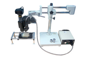
C. Boom Stand Microscopes
A boom stand microscope, also known as a stereo microscope with a boom arm or boom stand, can be a valuable tool for imaging microfluidic devices. While typically used for macroscopic observations, a boom stand microscope can also be adapted for microfluidics with certain considerations. Here are some key points to consider when using a boom stand microscope for microfluidics:
- Working Distance: One advantage of a boom stand microscope is its long working distance, which refers to the distance between the objective lens and the sample. This feature allows ample space for positioning microfluidic devices and facilitates easy access to the channels or features of interest.
- Macroscopic Observations: Boom stand microscopes are primarily designed for observing larger objects, such as electronic components or larger biological samples. Therefore, they are particularly useful for macroscopic observations of microfluidic devices, allowing for a broader view of the overall device, channel layouts, and fluidic connections.
- Sample Manipulation: The boom arm of the microscope provides flexibility and maneuverability, enabling the positioning and manipulation of microfluidic devices. This can be beneficial when aligning the device, introducing fluids, or adjusting the experimental setup during the imaging process.
- Magnification and Resolution: While boom stand microscopes offer lower magnification options compared to high-power compound microscopes, they can still provide sufficient magnification for microfluidic imaging, especially for larger structures within the device. However, for higher-resolution imaging or the visualization of smaller features or particles, additional magnification tools or objectives may be required.
- Imaging Technique Compatibility: Boom stand microscopes are primarily equipped with brightfield or reflected illumination systems. While these are suitable for observing larger structures and the overall device, they may have limitations when it comes to specific imaging techniques such as fluorescence or phase contrast. Additional modifications or attachments may be necessary to incorporate these techniques into the boom stand microscope setup.
- Sample Stability: It’s essential to ensure the stability of the microfluidic device during imaging. Boom stand microscopes may not offer the same level of stability as specialized microscopy setups. Consider using vibration isolation devices or securing the microfluidic device to minimize movement and vibration artifacts.
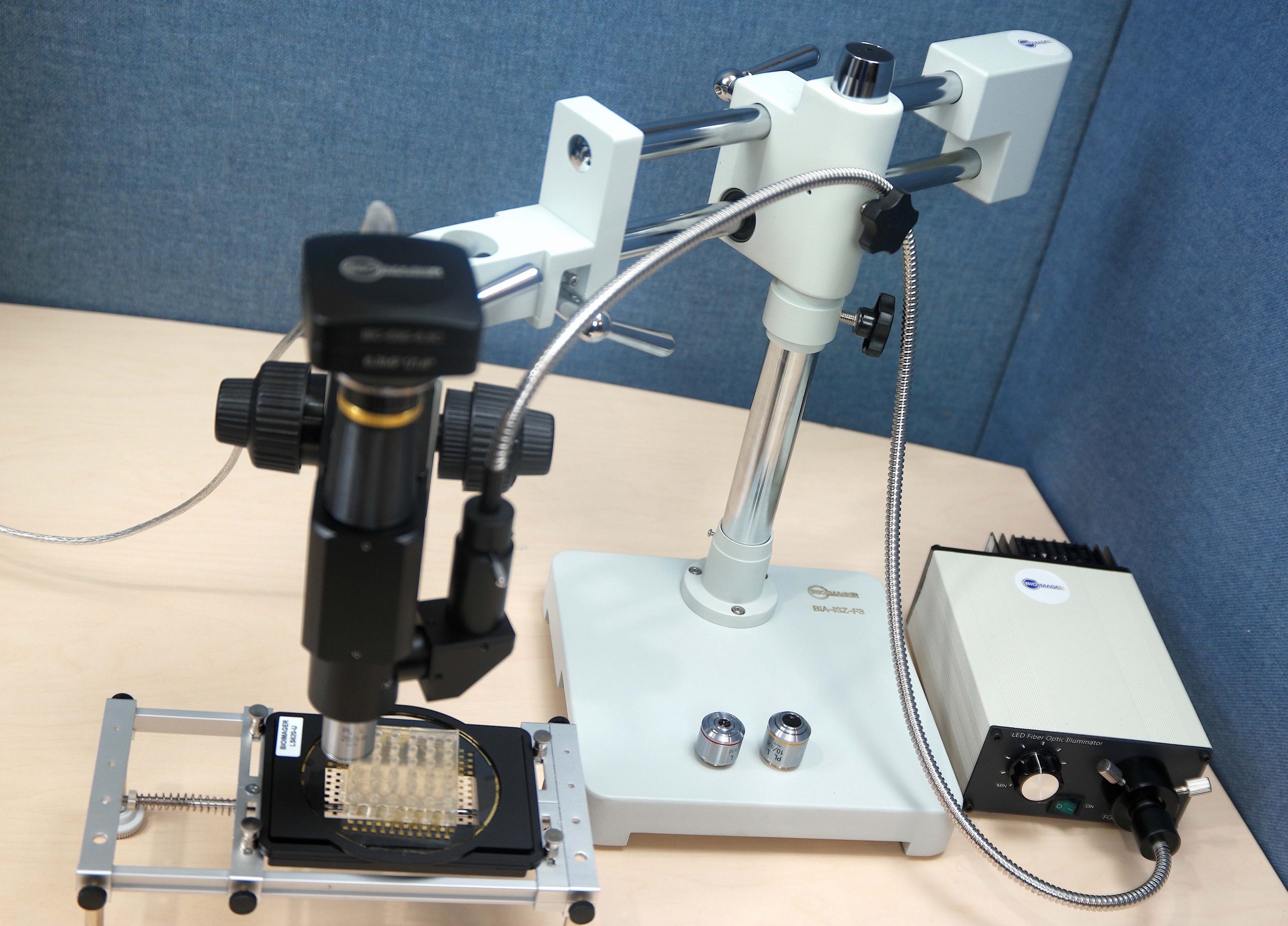
Client’s StoryIn this specific case, the client requested a movable inspection video microscopy system to visualize some phenomena occur inside a microfluidic chip. The observation needs to be done from top, i.e. an upright microscope is required. In this case a mechanical XY stage moves the device in X, or Y direction and lifts up/down the whole device objective to bring the image in focus. |
Custom Inspection Video Microscope with High Magnification
|
D. High Speed Camera
High-speed cameras are commonly used in microfluidics microscopy imaging to capture rapid processes and dynamic events within microfluidic devices. These cameras offer high temporal resolution, allowing for detailed analysis of fluid flow, droplet formation, particle behavior, and other dynamic phenomena. Here are some key points to consider when using a high-speed camera for microfluidics microscopy imaging:
- Frame Rate and Temporal Resolution: High-speed cameras are characterized by their frame rate, which represents the number of images captured per second. The frame rate determines the temporal resolution and the ability to capture fast processes. For microfluidic imaging, cameras with frame rates in the range of hundreds to thousands of frames per second (fps) are commonly used to capture dynamic events within microchannels.
- Shutter Speed and Exposure Time: High-speed cameras often have adjustable shutter speeds or exposure times, allowing for precise control over the exposure duration. This feature is crucial when imaging fast-moving objects or events within microfluidic channels. By adjusting the exposure time, researchers can prevent motion blur and capture crisp images of dynamic processes.
- Sensitivity and Low Light Performance: Microfluidic systems may involve low light levels, especially when imaging fluorescence or bioluminescence-based assays. High-speed cameras with high sensitivity and low light performance are desirable for capturing such low light signals. Consider cameras with low noise levels and high quantum efficiency for optimal imaging in low light conditions.
- Spatial Resolution: While high-speed cameras excel in temporal resolution, it’s important to consider their spatial resolution as well. The resolution determines the level of detail captured in the image. Select a camera with sufficient spatial resolution to observe the desired features or particles within the microfluidic channels.
- Triggering and Synchronization: To capture specific events or synchronize the camera with other devices or equipment, look for high-speed cameras that offer triggering and synchronization capabilities. This allows for precise timing and coordination with external triggers or events within the microfluidic system, ensuring the accurate capture of desired phenomena.
- Data Transfer and Storage: High-speed cameras generate a significant amount of data due to their fast frame rates. Consider the camera’s data transfer capabilities and storage options to ensure smooth data acquisition and storage during imaging experiments. Some cameras offer onboard memory or direct data transfer to a computer for efficient data handling.
- Integration with Microscope Setup: Ensure that the high-speed camera can be seamlessly integrated into your microscope setup. Consider the camera’s compatibility with the microscope’s imaging port, mounting options, and control interfaces. Consult with the camera manufacturer or microscope provider to ensure compatibility and proper integration.
E. Examples of our customers
Here is a list of some of our customers in microfluidic field that has used at least one of our products:
Canada: 1.University of Toronto, 2. McGill University, 3. York University, 4. McMaster University
USA: 1. Illinois University, 2. Stanford University, 3. Berkley University
When selecting a microscope for imaging microfluidics MEMS, consider the specific imaging requirements, such as resolution, imaging depth, surface characterization, and the need for additional imaging modalities. It’s also important to ensure compatibility with the size and configuration of the microfluidic MEMS devices being studied. Consulting with experts in the field or microscopy facilities can provide further guidance tailored to your specific experimental needs.