| Item |
Specification |
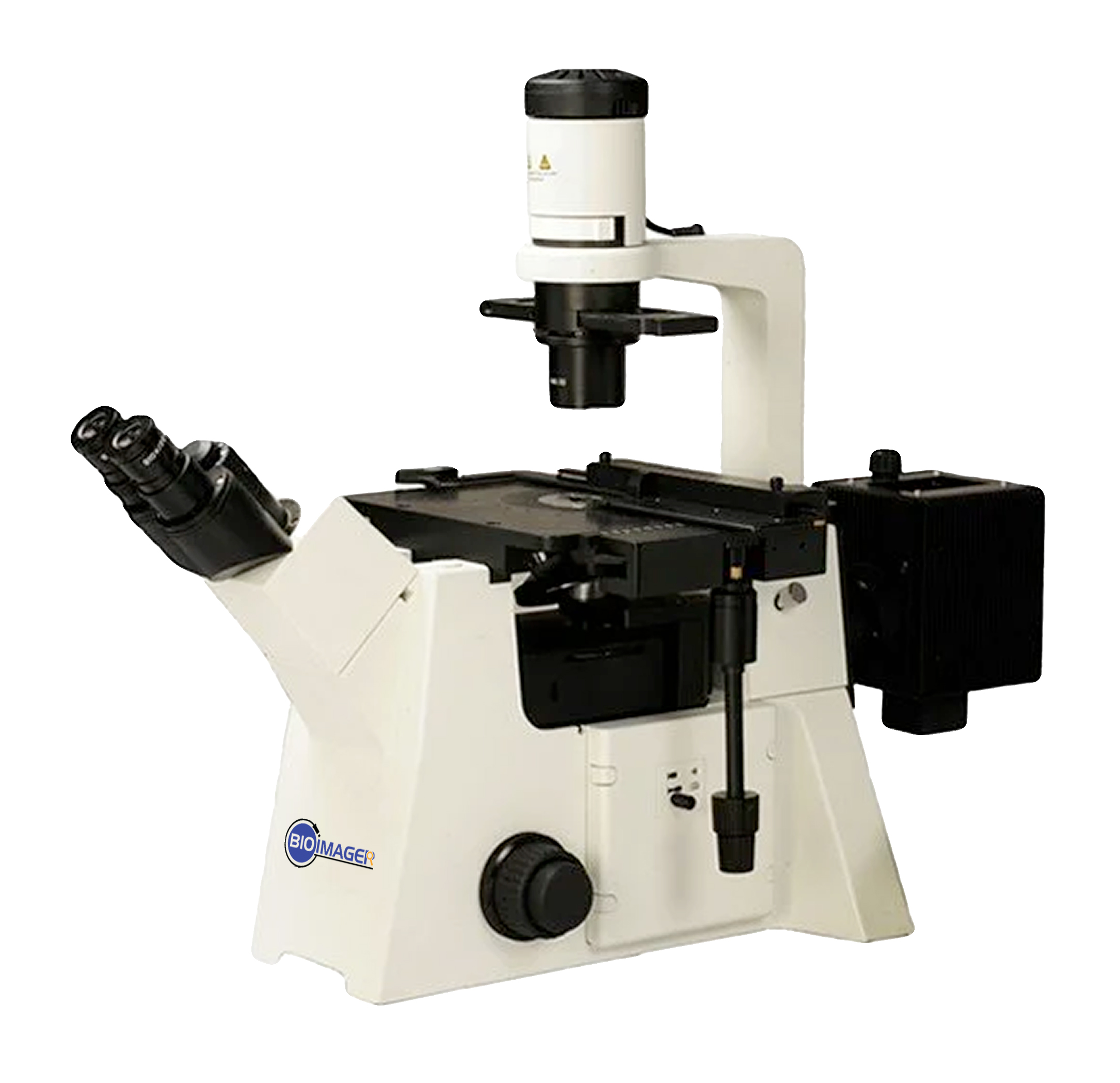
BIM630 |
BIM630FL |
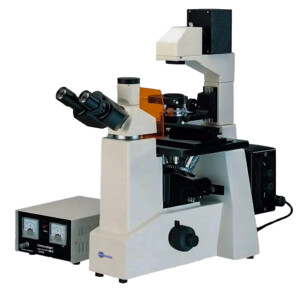
| Optical System |
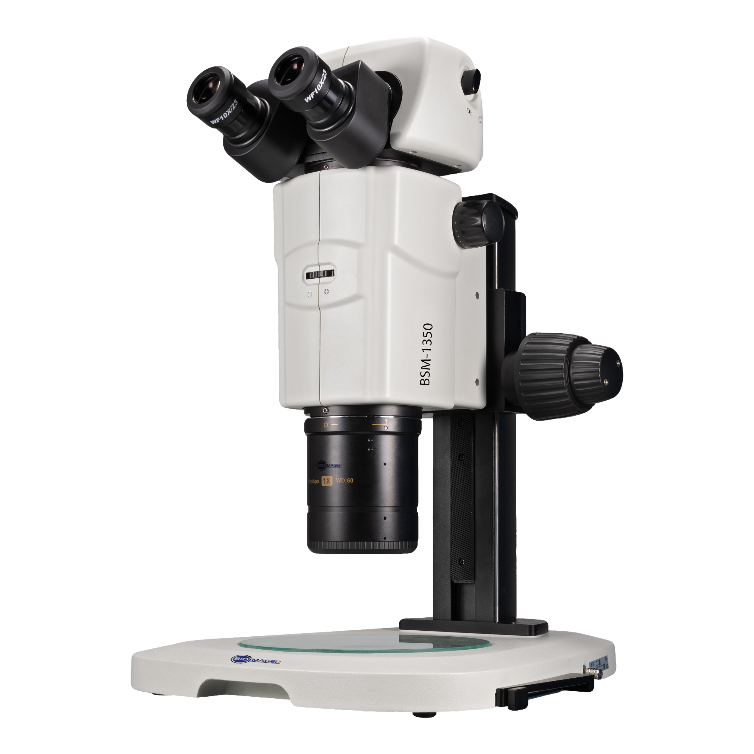
Focal length of 60mm Infinite Optical System, Tube length 200mm |
● |
● |
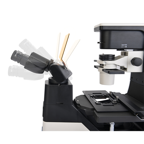
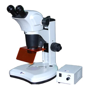
| Viewing Head |
Seidentopf Tilting Binocular Head, adjustable 5-35° inclined, Interpupillary Distance 48-75mm, Left side camera port, Light distribution: 100: 0 (100% for eyepiece), 0:100 (100% for camera), Eyepiece Tube Diameter 30mm |
● |
● |
| Eyepiece |
SW10×/ 22mm |
● |
● |
| WF15×/ 16mm |
○ |
○ |
| WF20×/ 12mm |
○ |
○ |
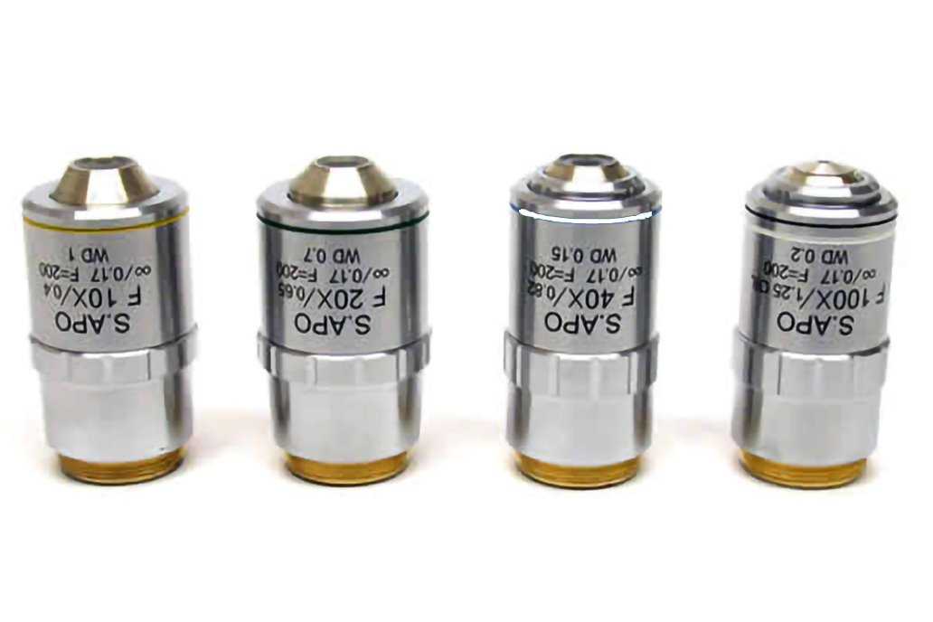
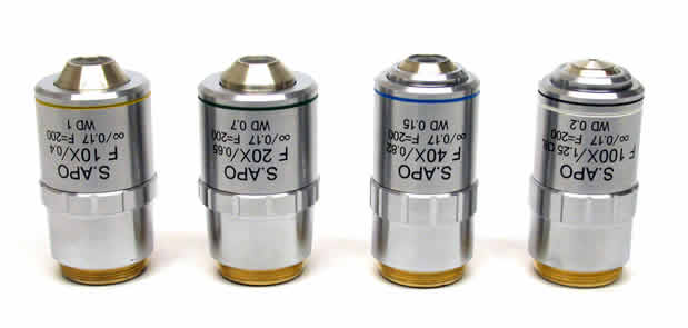
| Objective (Parfocal distance 60mm, M25×0.75) |
FL60 Infinite LWD Plan Achromatic Objective |
4×/0.1, WD=30mm |
● |
○ |
| 10×/0.25, WD=10.2mm |
○ |
○ |
| 20×/0.40, WD=12mm |
○ |
○ |
| 40×/0.60, WD=2.2mm |
○ |
○ |
| FL60 Infinite LWD Plan Phase Contrast Achromatic Objective |
PH10×/0.25, WD=10.2mm |
● |
○ |
| PH20×/0.40, WD=12mm |
● |
○ |
| PH40×/0.60, WD=2.2mm |
● |
○ |
| FL60 Infinite LWD Plan Semi-APO Fluorescent Objective |
4×/0.13, WD=17mm, cover glass=- |
○ |
● |
| 10×/0.3, WD=7.4mm, cover glass=1.2mm |
○ |
● |
| 20×/0.45, WD=8mm, cover glass=1.2mm |
○ |
● |
| 40×/0.60, WD=3.3mm, cover glass=1.2mm |
○ |
● |
| 60×/0.70, WD=1.8-2.6mm, cover glass=0.1-1.3mm |
○ |
○ |
| FL60 Infinite LWD Plan Semi-APO Phase Contrast Objective |
4×/0.13, WD=17.78mm, cover glass=- |
○ |
○ |
| 10×/0.3, WD=7.4mm, cover glass=1.2mm |
○ |
○ |
| 20×/0.45, WD=7.5-8.8mm, cover glass=1.2mm |
○ |
○ |
| 40×/0.60, WD=3-3.4mm, cover glass=1.2mm |
○ |
○ |
| 60×/0.70, WD=1.8-2.6mm, cover glass=0.1-1.3mm |
○ |
○ |
| Nosepiece |
Coded Quintuple Nosepiece |
● |
● |
| Condenser |
N.A. 0.3 Insert Plate Condenser, Working Distance 75mm |
● |
● |
| N.A. 0.4 Insert Plate Condenser, Working Distance 45mm |
○ |
○ |
| Telescope |
Centering Telescope: used to adjust the center of phase annulus |
● |
● |
| Phase Annulus |
10×-20×-40× Phase Annulus Plate (center adjustable) |
● |
● |
| 4× Phase Annulus Plate |
○ |
○ |
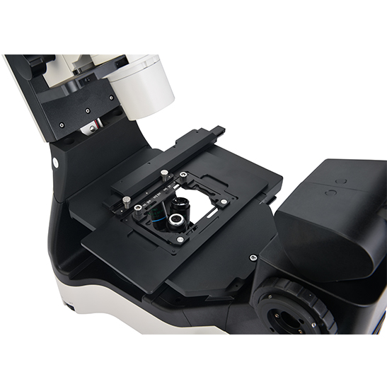
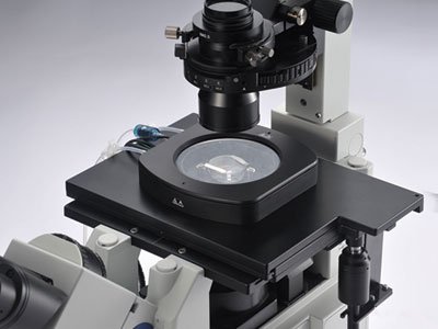
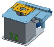
| Stage |
Stage 170 (X)×250(Y) mm with glass insert plate (diameter 110mm) |
● |
● |
| Attachable Mechanical Stage, X-Y Coaxial Control, Moving Rang: 128mm×80mm, accept 5 types of petri-dish holders, well plates and stage clips |
● |
● |
| Auxiliary stage 70mm×180mm, used to extend the stage |
○ |
○ |
| Universal Holder: used for Terasaki plate, glass slide and Φ35-65mm petri dishes |
● |
● |
| Terasaki Holder: used for Φ35mm Petri Dish Holder and Φ65mm petri dishes |
○ |
○ |
| Glass Slide and Petri Dish Holder Φ54mm |
○ |
○ |
| Glass Slide and Petri Dish Holder Φ65mm |
○ |
○ |
| Petri Dish Holder Φ35mm |
○ |
○ |
| Petri Dish Holder Φ90mm |
○ |
○ |
| Focusing |
Coaxial Coarse and Fine Adjustment, tension adjustment, Fine Division 0.001mm, Fine stroke 0.2mm per rotation, Coarse stroke 37.5mm per rotation. Moving Range: up 7mm, down 1.5mm; Without limitation can up to 18.5mm |
● |
● |
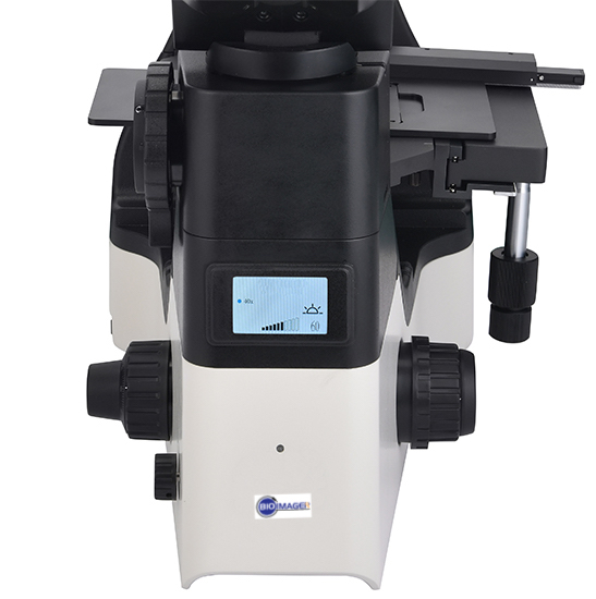
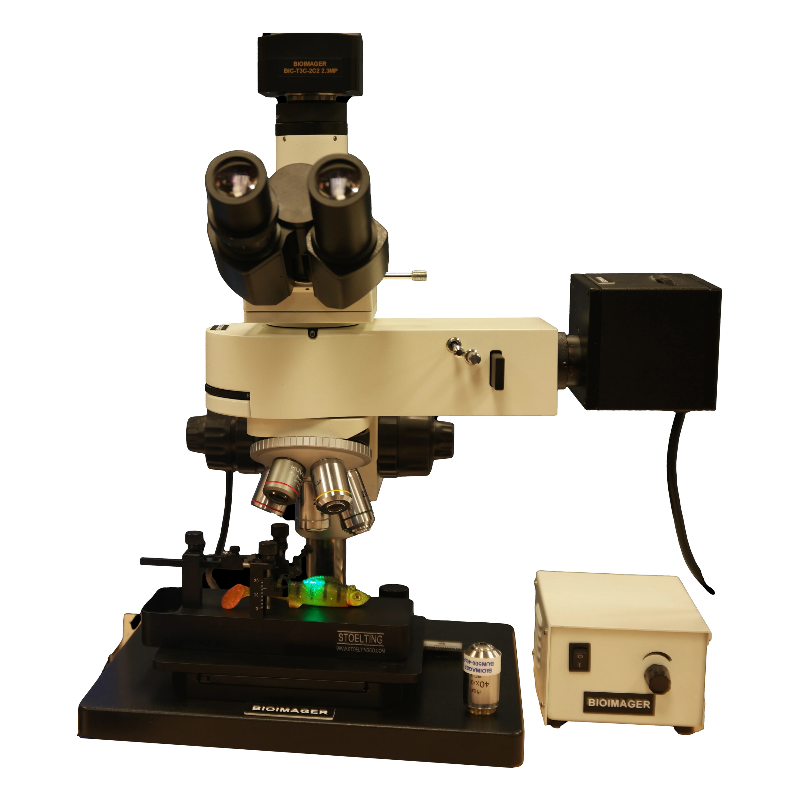
| Transmitted Illumination |
3W S-LED Koehler illumination, Brightness Adjustable |
● |
● |
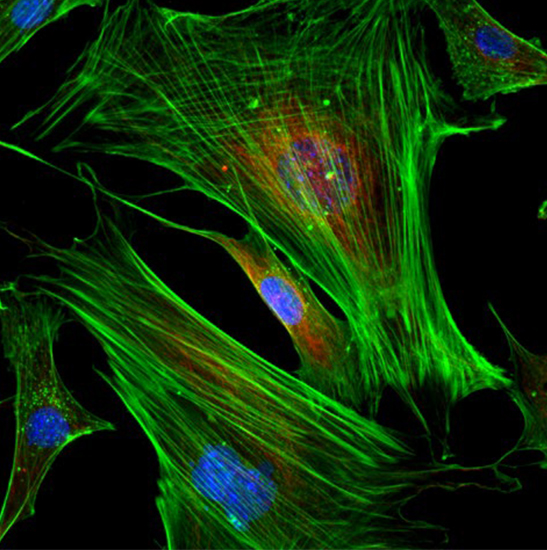
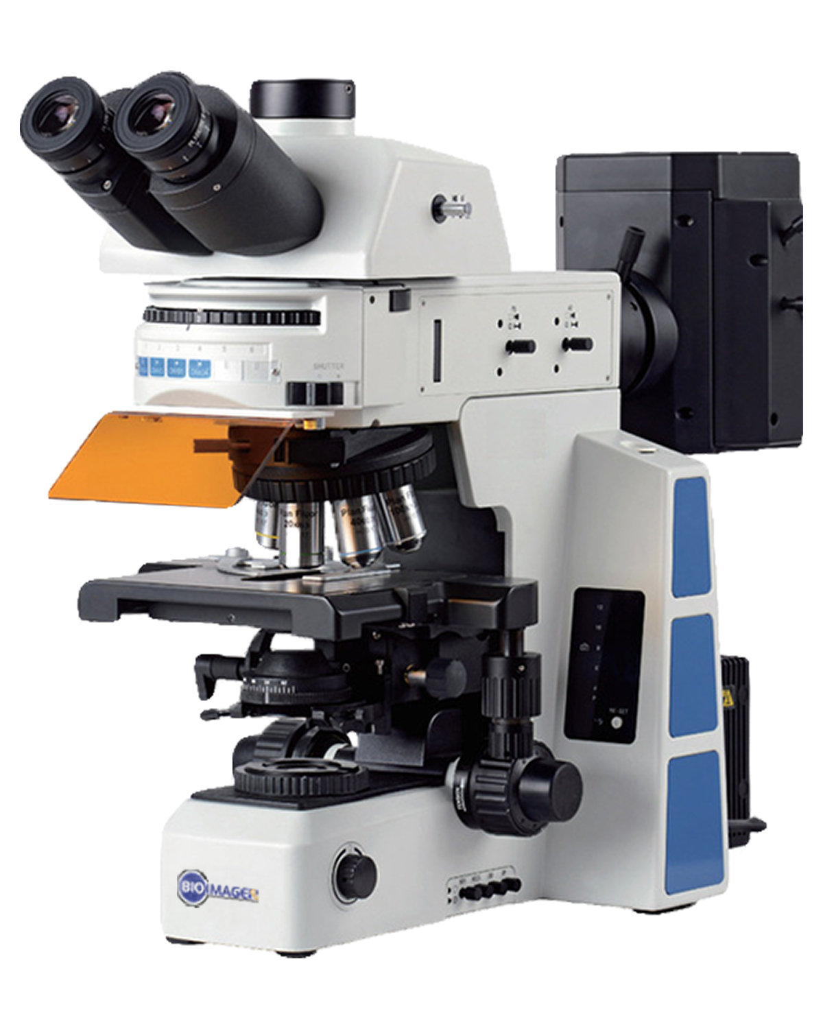
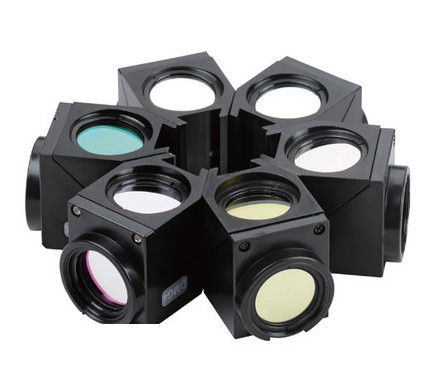
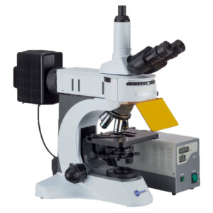
| EPI-Fluorescent Attachment |
LED illuminator, built-in Fly-eye lens, can be configured with up to 3 different LED light source and B, G, U fluorescent filter blocks |
○ |
● |
| LED light source and V, R, FITC, DAPI, TRITC, Auramine, mCherry fluorescent filters |
○ |
○ |
| Hoffman phase contrast |
Hoffman Condenser with 10×, 20×, 40× insert plate, centering telescope and special objective 10×, 20×, 40× |
○ |
○ |
| 3D Emboss Contrast |
Main emboss contrast plate with 10×-20×-40× will be inserted into condenser |
○ |
○ |
| Auxiliary emboss contrast plate will be inserted into slot near viewing head |
○ |
○ |
| C-mount Adapter |
0.5× C-mount Adapter (focus adjustable) |
○ |
○ |
| 1× C-mount Adapter (focus adjustable) |
● |
● |
| Other Accessories |
Warm stage |
○ |
○ |
| Light shutter, can be used to block the external light |
○ |
○ |
| Dust cover |
● |
● |
| Power Supply |
AC 100-240V, 50/60Hz |
● |
● |
| Fuse |
T250V500mA |
● |
● |
| Packing |
2cartons/set, Packing Size: 47cm×37cm×39cm, 69cm×39cm×64cm, Gross Weight: 20kgs, Net Weight: 18kgs |
● |
● |
Reviews
There are no reviews yet.