Slide Scanners

Showing 1–9 of 17 results
-
-

-
 Request a Quote Request a Quote
 -
-
Request a Quote Request a Quote
-
-
$USD 1,495.00 Add to cart
-
-
Showing 1–9 of 17 results
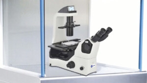
Digital Slide Scanning System
A) For pathology, histology and biology application
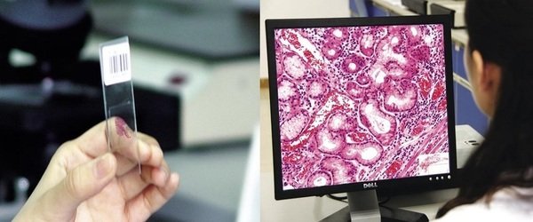
A Microscope Slide Scanner, also known as Digital Pathology Scanner or Digital Histology Slide Scanner, produces quick, reliable and high-resolution images of a glass slide. Pathologists, histologists, biologists, hematologists, vet experts or medical professionals now have the ability to scan single or often multiple slides. They can upload these images onto a network, iCloud store for remote access and collaboration among colleagues immediately or within a few minutes. Features may include remote control, real-time viewing, sharing, commenting, measurements, expert notes, reports, and many advanced software integration.
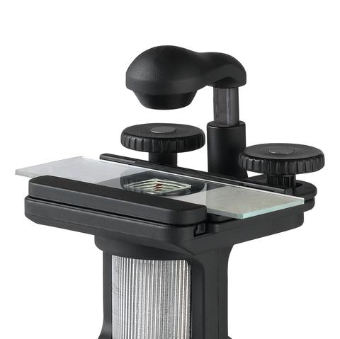
A Digital Pathology Scanning System may provide automated tissue slide imaging, counting and measurement for both fixed or live-cell assays. A microscopy system for slide scanning have modes for fluorescence, phase-contrast, polarizing and darkfield imaging. All slide scanners work for a standard size 1″x3″ (25x75mm) and some for double size 2″x3″ (50mm x75mm) , either glass or plastic slides with/without coverslips.
Digital slide scanners are widely used in pathology, histology, and biology applications for the purpose of digital imaging and analysis of tissue samples. Here are some factors to consider when choosing a digital slide scanner for these applications:
- Resolution: A high-resolution scanner is important for accurately capturing the details of tissue samples. A resolution of 20x or higher is generally recommended for pathology applications.
- Scan speed: The scan speed of the scanner is important to consider, especially for labs with a high volume of samples. Faster scanning times can improve efficiency and throughput.
- Image quality: The quality of the scanned images is crucial for accurate analysis and diagnosis. Look for a scanner that produces clear, high-quality images with accurate color representation.
- Compatibility: Make sure the scanner is compatible with your existing software and hardware. Some scanners may require specific software or hardware to function properly.
- User-friendliness: Consider the ease of use of the scanner and its software. A user-friendly interface can save time and reduce errors during operation.
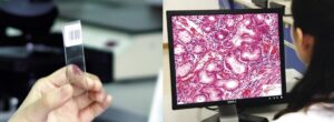
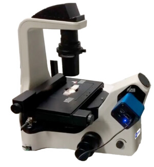
A digital slide scanning system is a specialized microscope that captures high-resolution digital images of whole microscope slides for pathology, histology, and biology applications. This technology allows researchers and clinicians to view and analyze virtual slides on a computer screen, instead of using a traditional microscope.
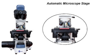
The system typically includes a high-resolution camera, motorized stage, and advanced imaging software that allows for rapid and precise scanning of entire slides. The resulting digital images can be viewed, analyzed, and shared with other researchers and clinicians for collaboration and diagnosis.
Digital slide scanning systems are commonly used in pathology for the diagnosis of cancer and other diseases. They enable pathologists to view and analyze digital slides remotely, which can be particularly useful in situations where specialists are not located in the same geographical area as the patient.
In addition to pathology, digital slide scanning systems are also used in histology and biology applications. Researchers can use the technology to capture high-quality images of tissue samples, which can be used for a variety of research purposes, including investigating disease mechanisms, developing new treatments, and studying the effects of drugs on tissue samples.
Overall, digital slide scanning systems are powerful tools for pathology, histology, and biology applications, allowing for high-resolution imaging and analysis of tissues and cells. They are widely used in research labs, hospitals, and clinics around the world.
There are so many digital slide scanner brands for pathology, histology, and biology applications in the market. It’s important to carefully evaluate your specific needs and choose a scanner that best fits your requirements.
B) For Mineralogy and Geology Applications
Slide scanning system for mineralogy or geology slides is very similar to that of pathology slide scanner. It comes with a polarizer and rotating analyzer. Analyzers can be rotated from 0 to 90 degrees (and often to 360 degrees)manually or fully control with the software. A mineralogy slide scanner may have both transited and reflected lights while a pathology slide scanner uses transmitted light only.
7 Things I wish I know before buying a slide scanner:
- Slide Size: The very first and important thing about a slide scanner is the size of slide. Some scanners accepts only standard size of 25mm x 75mm (or 1″ x 3″), while few models of scanners can also get the double size slides which are 50mm x 75mm (2″ x 3″).
- A Number of Slides: A regular slide scanner obviously can read one slide, but it happens clinical labs or hospitals deal with tens of hundreds of slides every day. Do I need an automated slide loader? The answer depends on the number of slides per day and of course budget. Some scanners also offer barcode reader.
- Scanning area: a standard coverslip for a standard size glass slide is 20mm x 50mm (0.8in x 2in). Do you need to scan the whole cover-slip size or just a small fraction of that?
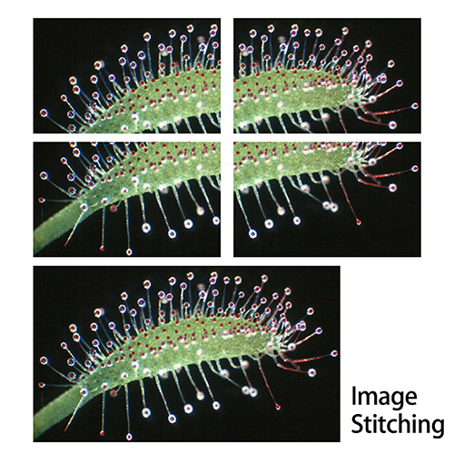
- Scanning speed: This can be the most important question when you look for a new slide scanner. Bu the answer is not simple. This depends on few factors such as the scanning area size, magnification (or objective lens), and price. Some models of scanners (such as uScope) offers two cameras, one is a webcam which gives the whole slide view and the second camera comes from the objective lens. The speed can also vary depending on the z-stacking to be done while scanning or not.
- Z-stacking: everyone has own preference when he or she looks at a 3D image or particle. When you use over 20x magnifications, using a single Z plane is not enough, thus Z-stacking is required. Most of the time we do not need to do Z-stacking on all over the slide area but limited ROI (regions of interest) is enough. You can use special software for this purpose.
- Price: This might be as #1 priority in list for some of the customers. As you know well, the price is not everything. However, the budget is the question you know its answer better. One thing we, Bioimager, can add in this regard is that if you need to scan at multiple magnifications, you may be better not choose an expensive scanner which can give you multiple magnification option but simple buy two low- or medium-price slide scanners with different magnifications.
- Can I convert my current microscope to a digital slide scanner? Yes, you can. Simply check our software options among the products. BioStitch-500 and BioStitch-1000 are made exactly for these purpose.
Our Customers:
Here are examples of customers who have tried our slide scanners: