Stereo Microscopes
Showing 1–9 of 59 resultsSorted by latest
-
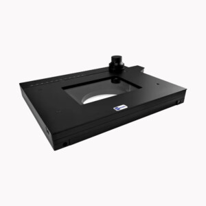
BSM-XY-284 Microscopy Stage high quality Large travel distance 110*110mm XY Working microscope stageOriginal price was: $USD 2,000.00.$USD 1,280.00Current price is: $USD 1,280.00. Add to cart
Showing 1–9 of 59 resultsSorted by latest
Stereo Microscopes
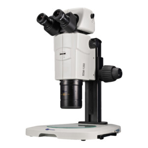
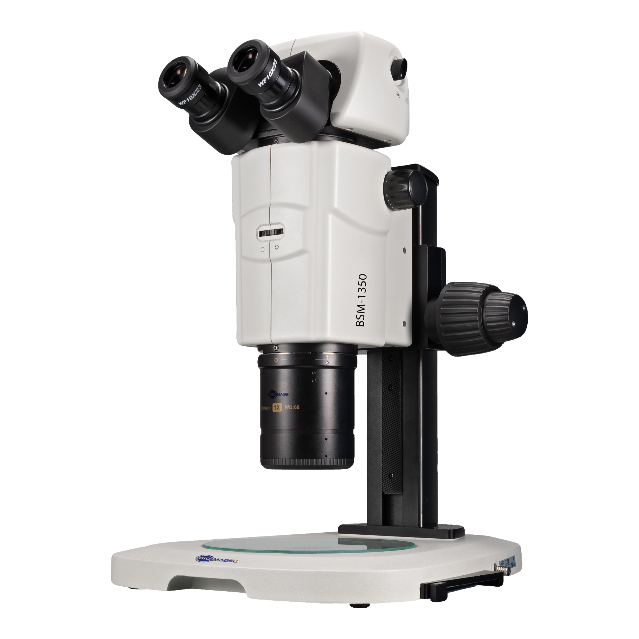
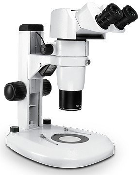
A stereo microscope, also known as a stereoscopic microscope or a dissecting microscope, is a type of microscope used for the examination of objects that are too large or thick to be observed under a conventional compound microscope.
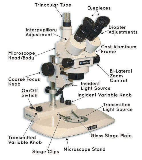
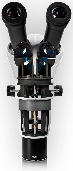
A stereo microscope has two separate optical paths that provide a three-dimensional image of the sample, giving a stereoscopic or binocular view. This is achieved by using two separate objective lenses and two eyepieces, each with its own optical pathway.
Unlike a compound microscope, a stereo microscope has a low magnification power and provides a wider field of view, making it suitable for examining larger specimens. It is commonly used in biology, botany, entomology, forensic science, and other fields that require the examination of larger specimens, such as insects, plants, rocks, and small electronic components.
Stereo microscopes may also have additional features such as a zoom function to adjust the magnification, built-in illumination to provide better visibility of the sample, and a range of interchangeable lenses to provide different magnification options.
Overall, stereo microscopes are valuable tools for examining and studying larger specimens and are widely used in various scientific fields.
| Stereo microscopes are low power microscopes which are also called dissecting, dissection or inspection microscopes. Stereoscopes have low power or magnification range which is mainly 10x-70x (10-70 times bigger than actual).
Stereo microscopes are used for inspection and visualization of stamps, coins, mobile parts, printed circuit board (PCB), insects, plants, seeds, fruits, veterinary, anatomy, nearoanatomy, zebrafish, in vitro fertilization (IVF), music instrument parts (strings, CDs,…), material surfaces, ….. . Adding a polarization kit, stereoscopes can be used for inspection of rocks, minerals, and fossils. Advanced studies in this regard, which requires higher magnification, can use Polarizing microscopes or metallurgical microscopes. Researchers in zebrafish, embryo, IVF, bacteriophage, and microfluidics on small animal models like c-elegant use fluorescence stereoscopes. Using auxiliary (or aux) lenses, you can extend the magnification or field of view. With a 2x aux lens, 30x eyepieces, in a regular stereoscope (0.7x-5.6x), the max magnification can be 336x. If a smaller magnification aux lens such as 0.5x or 0.33x is used, the working distance (WD) and field of view (FOV) will be increased and you can expect to see as large as 50mm FOV and over 200mm WD. Contact us for such a configuration. By default, all light microscopes generate brightfield images, i.e. specimen is seen with a bright background. It is possible to see the images with a dark background, i.e. darkfield images. To compare this, think of seeing an airplane in the sky during the day (brightfield) versus seeing it near sunset (darkfield). You can add a darkfield and polarization kit to a stereoscope to make it much professional. Contact us for further details. What to consider before buying: Magnification: a typical magnification is 7x-45x which can be extended using a 15x, 20x, 30x eyepieces and /or 1.5x / 2x aux lenses Working distance: at physical stereoscope has 80-90mm WD. illumination: Reflected for opaque sample and transmitted light for transparent samples. |
 |
Case Study: The larger field of view, the better for teaching
A client wrote us: Interested in a trinocular zoom stereoscope with the widest field of view (to the camera, we have a Lumenera Infinity2-1R, 1/2″ 1.4 MPixel) possible. Probably would need 0.5X aux lens. BUT CAN YOU PROVIDE for me with the field-of-view we could achieve (with our camera)? We need to use this to teach neuroanatomy, meaning about 2 cm horizontal field of view (at lowest mag setting).
Solution:
To get the largest field of view (FOV), a good combination of the camera sensor and the c-mount adapter is the key. As you know well, we see circular in eyepieces but a camera sees a rectangular or square. Depending on the c-mount adapter magnification, we can fit the camera FOV square or rectangle into the eyepiece circle. If we go further than eyepiece FOV circle, there won’t be any signal to the corners of the square. Then the sensor will generate an image with black corners (called vignetting).
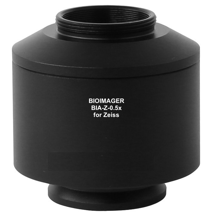
Depending on the stereoscope model, it is possible to get ~20-22mm width image assuming a perfect camera-adapter is used. In most cases, we can reach this without a 0.5x aux lens. Since you like to use your own camera with 1/2” sensor, we need to provide a 0.5x c-mount adapter.
You need to use BSM330T or higher models to get such a large FOV. Using a 0.5x aux lens will be an advantage.
Next, we asked the client to send his own sample and camera to test. Here are the rest of further works:
We have done some works and sending you the images in a separate email via an external link. Please read these comments before and after viewing those images.
– The file name describes the imaging condition: date_camera_c-mount-adapter_scope zoom_sample-Name.jpg
– we tested so many c-mount adapters, including default and custom 0.35x, 0.5x and 1x c-mount adapter with your Infinity 2 Camera. Some of the images are presented.
– The 0.35x gives largest FOV but it has some vignetting issue, as expected.
– The custom 0.5x c-mount adapter gives a better result with your camera. However, it has a minor black corner on one side. You may not like it.
– The 1x c-mount adapter helps you increase the magnification twice in comparison to 0.5x c-mount adapter, i.e. it will be like using 20x/FN10 eyepieces vs 10x /FN20 or adding a 2x aux lens.
– At 0.85x zoom, you can have the whole view of your sample
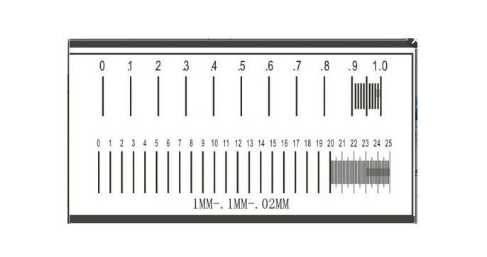
– Using a calibration slide, now we know your camera has an FOV of 20mmx15mm (HxV) at the lowest magnification 0.7x zoom.
If you do not like to have black corners in your images, you need to either upgrade the microscope or change the camera. This will depend on your budget.
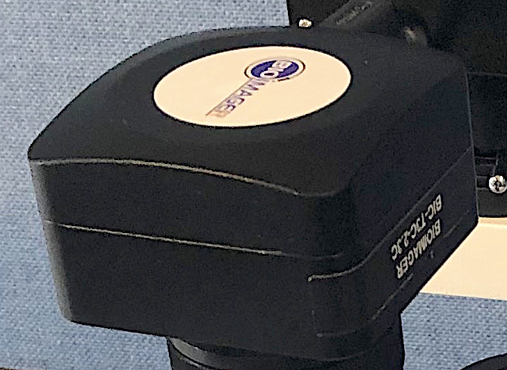
Thus, I tested this with our BIC-T3C-2.3C 2.3MP 1/1.2” CMOS camera.
– This camera has an FOV of 22.5x14mm (HxV) at the lowest magnification 0.7x zoom.
– Some sample images were also shown.
– Frankly speaking, this camera has better image quality, much more natural color and higher sensitivity than yours. I guess ours has also better price, i.e. $1,089 USD.
Last but not least, we also used our BioStitch Live Image Stitching Software and manually stitched your sample images at the highest zoom of 4.5x and 0.5x c-mount adapter. Using 1x c-mount adapter will increase the resolution twice. If you decide to get the BIC-T3C-2.3C camera, we can offer this software ($1,499) totally free.
Result: the customer was extremely happy with the prepared images, purchased the stereoscope and enjoyed it.
Stereo Microscopes: Tips and Tricks
Thanks Brian Kloepfer, Carolina Biological Supply Co.
Please select the products based on your requirements from the below table:
