Illumination
Showing 1–9 of 17 resultsSorted by latest
Showing 1–9 of 17 resultsSorted by latest
Illumination for Microscopy Imaging
Microscopy illumination refers to the source of light used to illuminate biological samples in microscopy. The choice of illumination depends on the type of microscopy, the nature of the sample, and the desired imaging outcomes.
There are several types of microscopy illumination, including brightfield, darkfield, phase contrast, and fluorescence.
Brightfield illumination is the most common type of illumination and involves the use of white light to illuminate the sample. The sample appears dark against a bright background, making it easy to observe the morphology of cells and tissues.
Darkfield illumination is similar to brightfield illumination, but it involves the use of oblique light that is not collected by the objective lens. This results in a bright image of the sample against a dark background, making it useful for visualizing transparent or unstained samples.
Phase contrast illumination is used to enhance the contrast of transparent samples, such as live cells or tissues. It involves the use of a phase ring in the condenser to create a phase shift in the light passing through the sample, resulting in an image with high contrast.
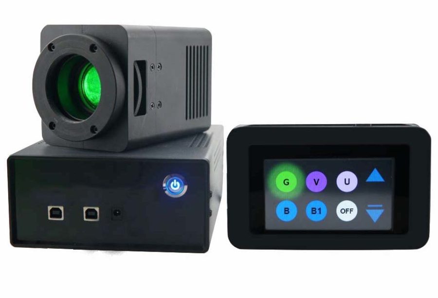
Fluorescence illumination involves the use of specific wavelengths of light to excite fluorescent dyes or proteins in the sample. The excited fluorophores emit light at a different wavelength, which is captured by a camera and used to generate a fluorescence image. This type of illumination is useful for visualizing specific molecules or structures within cells and tissues.
Other types of microscopy illumination include polarization microscopy, differential interference contrast (DIC) microscopy, and confocal microscopy. The choice of illumination depends on the specific imaging requirements of the experiment.

