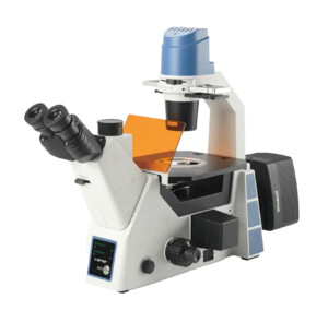
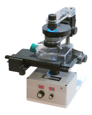
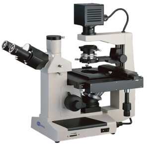
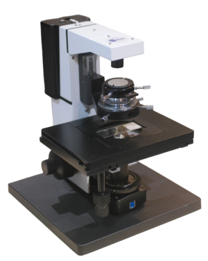
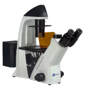
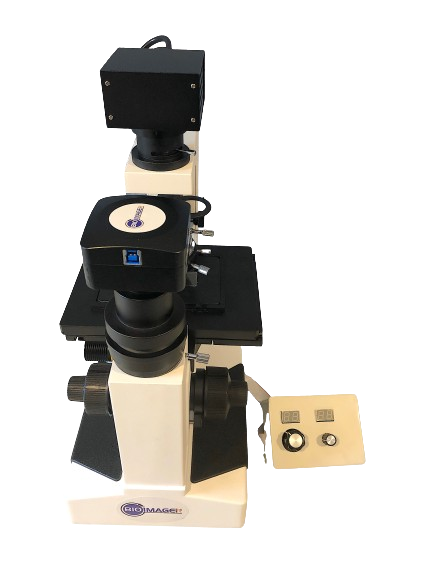
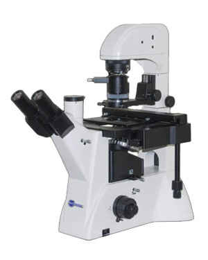
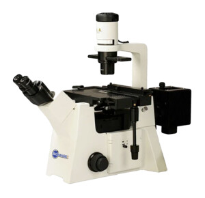
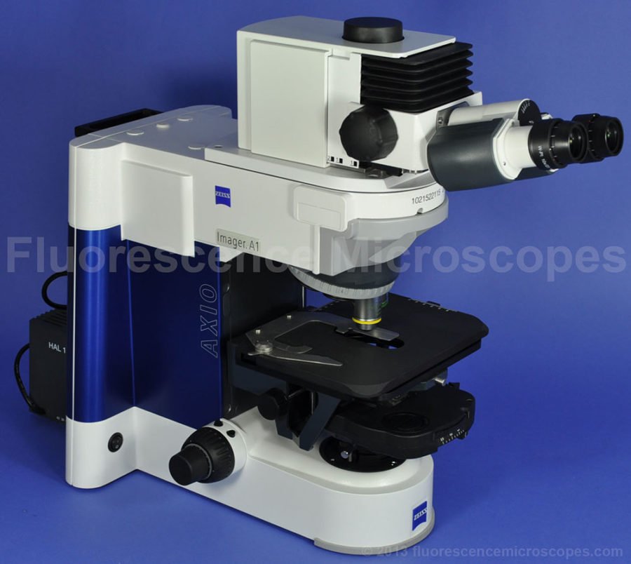
Phase Contrast Microscopes

Showing 1–9 of 25 results
-
-
-
 Request a Quote Request a Quote
 -
$USD 3,995.00 Add to cart
-
$USD 9,750.00 Add to cart
-
-
-
-
$USD 14,000.00 Add to cart
Showing 1–9 of 25 results
Introduction to Phase Contrast Microscopy Imaging
Phase contrast microscopes are commonly used in biological and medical research to observe transparent and unstained specimens, such as living cells or bacteria. Here are some factors to consider when choosing a phase contrast microscope:
- Objective lenses: The objective lenses are crucial for producing high-quality images of transparent specimens. Look for microscope objectives that have a high numerical aperture (NA) and are specifically designed for phase contrast microscopy.
- Condenser: The condenser is important for directing light onto the specimen and producing contrast. Look for a microscope with a phase contrast condenser that matches the objective lenses.
- Magnification range: A phase contrast microscope should have a wide range of magnification options to accommodate different types of specimens and applications. Look for a microscope with a magnification range of at least 10x to 100x.
- Illumination: The microscope should have adjustable illumination options to ensure proper lighting for different types of specimens. LED illumination is recommended for its long lifespan and low power consumption.
- Ergonomics: The design and ergonomics of the microscope should allow for comfortable, extended use without causing eye strain or discomfort. Look for microscopes with adjustable eyepieces and a comfortable working distance.
Several products of phase contrast microscopes are available to choose. It’s important to carefully evaluate your specific needs and choose a microscope that best fits your requirements. Additionally, it may be helpful to consult with experts in the field to determine the most appropriate microscope for your particular application.
How does a Phase Contrast Microscope Work?
A phase contrast microscope works by converting differences in refractive index within a specimen into contrast in the final image. This is particularly useful for observing transparent and unstained specimens, such as living cells or bacteria.
In a phase contrast microscope, light from the microscope’s illumination source passes through a phase plate, which is typically located in the condenser. The phase plate is designed to create a phase shift in the light passing through it, such that the light waves become slightly out of phase with one another.
Next, the light passes through the specimen, which causes further phase shifts depending on the specimen’s refractive index. The light waves then pass through the objective lens, which is also designed to create a phase shift.
Finally, the light waves pass through a second phase plate, which is typically located in the eyepiece. This phase plate recombines the light waves and produces an image with visible contrast between different regions of the specimen based on differences in refractive index.
By adjusting the alignment and position of the phase plates, it is possible to optimize the contrast and clarity of the final image. The result is an image with enhanced contrast and detail, even in specimens that would otherwise be difficult to observe using conventional brightfield microscopy.
History
The idea of Phase Contrast Imaging was brought by Frits Zernike in 1930’s, a Dutch physicist, who got the Nobel prize in 1953 for this invention.
Applications
Phase contrast is mainly used by biologists and has some advantages over brighfield imaging. Using phase contrast microscopy, many cellular structures that cannot be seen by a brightfield imaging become visible, without staining.
Summary
Phase Contrast is a contrast enhancing optical microscopy technique in which an unstained transparent sample is seen with high brightness by shifting phase difference of light.
Video Tutorial: Adjusting Phase