FISH Hybridization System
Showing the single result
Showing the single result
FISH Hybridization System
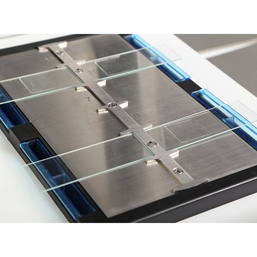
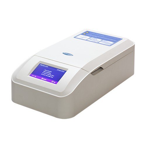
Fluorescence in-situ hybridization (FISH) is a technique used in molecular biology to detect and visualize the presence or absence of specific DNA or RNA sequences in chromosomes, cells, or tissues. FISH hybridization systems are laboratory instruments and reagent kits that enable researchers to perform FISH experiments.
The FISH hybridization system typically includes fluorescently-labeled DNA or RNA probes that are designed to hybridize with specific target sequences in the sample, as well as buffers, blocking agents, and other reagents that optimize the hybridization process. The system also includes a fluorescence microscope or imaging system that is used to visualize the hybridized probes and to analyze the results.
The FISH hybridization process involves several steps, including sample preparation, probe labeling and purification, hybridization, and signal detection. The sample is typically fixed and permeabilized to allow the probes to penetrate the cells or tissue. The probes are then labeled with fluorescent dyes and hybridized to the target sequences under optimized conditions. After hybridization, the excess probes are removed, and the sample is washed to remove any unbound probes. Finally, the sample is imaged using a fluorescence microscope or imaging system, and the results are analyzed to determine the presence or absence of the target sequences.
FISH hybridization systems have many applications in genetics and molecular biology, including gene mapping, chromosome analysis, gene expression analysis, and cancer diagnosis. They are widely used in research labs, clinical laboratories, and diagnostic centers.