Darkfield
Showing 1–9 of 32 results
-
-
-
-
-
-
-
$USD 11,920.00 Add to cart
-
-
Showing 1–9 of 32 results
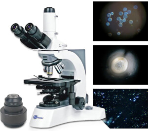
Darkfield Imaging
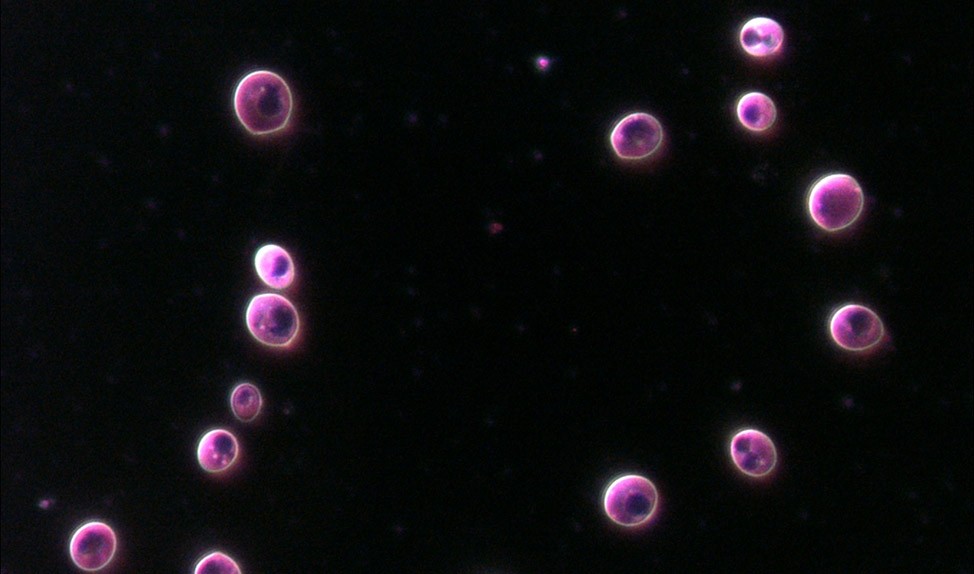
Darkfield microscopy is a specialized technique used in microscopy to enhance the visibility of transparent or low-contrast specimens. Unlike brightfield microscopy, where the specimen is illuminated directly, darkfield microscopy involves illuminating the sample with oblique or angled light, resulting in a dark background while the specimen appears bright.
In darkfield microscopy, a special darkfield condenser is used to direct the light at a specific angle so that it does not enter the objective lens directly. As a result, only the scattered light or light that interacts with the specimen is collected by the objective lens and observed.
The dark background produced by darkfield microscopy enhances the visibility of transparent or low-contrast specimens by increasing the contrast between the specimen and its surroundings. This makes it particularly useful for observing live, unstained samples, such as microorganisms, small organisms, and subcellular structures.
Darkfield microscopy is commonly used in various fields of study, including microbiology, cell biology, and materials science. Some of its applications include:
- Microorganism Observation: Darkfield microscopy allows for the visualization of bacteria, protozoa, and other microorganisms that may be difficult to see using other techniques. It can be useful for studying their morphology, motility, and interactions.
- Blood Cell Analysis: Darkfield microscopy can be employed to observe blood cells, including red blood cells, white blood cells, and platelets. It enables the examination of their structure, shape, and interactions.
- Transparent Organisms and Tissues: Darkfield microscopy is suitable for observing transparent organisms, such as aquatic organisms, larvae, and embryos. It can provide insights into their development, behavior, and interactions with their environment.
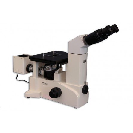
- Material Analysis: Darkfield microscopy is also used in materials science to examine the surface characteristics and defects of materials, such as nanoparticles, fibers, and thin films.
By providing a contrasting background, darkfield microscopy allows for the visualization of specimens that might be challenging to observe using other microscopy techniques. It offers a unique perspective and can reveal details and features that may otherwise go unnoticed.
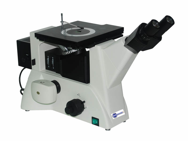
Darkfield imaging is very popular for imaging of live or deal blood cells, bacteria, virus and microorganisms particularly for those living in water. BIOIMAGER offers various type of microscopes with darkfield, dark-field, dark field imaging capabilities. This is for reflected (incident) or transmitted light in metallography or metallurgical microscopes and biological microscopes in either upright or inverted microscopes. These dissecting microscopes come with darkfield condensers and objective lenses. It is a useful technique for live blood cell imaging.
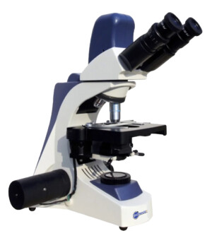
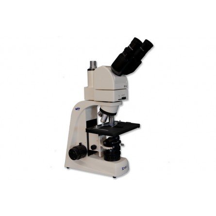
Darkfield illumination
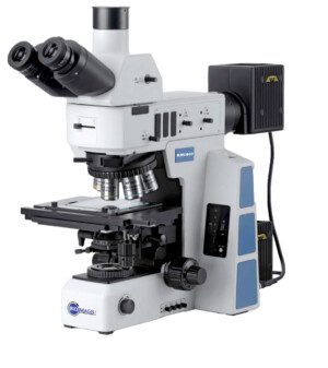
This short article concentrates on darkfield illumination. For this article, we will be using BUM500DF microscope. Darkfield illumination is a lighting method used to increase the contrast in low contrast samples. It is particularly useful for highlighting specific sample material while other methods such as phase contrast are better for viewing interior details. At the heart of dark field illumination there is a darkfield condenser. It works similarly to a brightfield condenser except it includes an opaque disc or stop which blocks direct light from entering the objective lens. Instead, only oblique lighting which is refracted by the sample will enter. This results in certain objects being illuminated while the background remains dark.
When choosing a dark field condenser be sure the condenser’s numerical aperture (NA) is larger than a numerical aperture of the objective lens you plan to use. If the objective lens’s aperture is larger than the condenser’s, a direct light will be transmitted and a dark field will not be achieved. At the dry condenser’s upper limit, an image shows noticeable glare. This is well above the condenser’s limit where the dark field is completely lost. By reducing the aperture of the lens, the dark field may be restored.
There are two classes of condenser dry and oil immersion. A dry condenser is designed to work with lower magnification objectives with lower numerical apertures. This typically covers objectives up to 40x (or may be 60x) with apertures of around 0.65 or less. An oil immersion condenser is needed for higher magnification lenses such as a 100x lens. Due to the high numerical aperture of these lenses, typically 1.25 or higher, a condenser of correspondingly high aperture is needed specially to maintain high resolution. while this combination will produce an image with improved contrast, it may not be truly darkfield due to the objective lenses high aperture. Thus, as an option a variable aperture lens may be used to fine tune and improve the darkfield image quality by closing a built-in iris diaphragm. The numerical aperture of the objective can be reduced until the field is adequately dark.
Replacing the condenser is a simple process. The condenser can be raised or lowered by using the adjustment knob. While supporting the installed condenser, loosen the locking screw until a condenser can easily slip out. Next, insert the darkfield condenser fully. Then, securely tighten the locking screw(s). A darkfield condenser should be centered for optimal results. Although the procedure is similar for dry and oil condensers, there are some minor differences for the dry condenser. Use a low powered objective preferably at 10x, by removing an eyepiece an image of the condensers lens can be seen. Adjust the height of the condenser and the stage (if necessary) until the opaque disc fills slightly less than the entire image circle leaving a ring of light. Now, use the two centering screws to move the disc until it appears centered within the ring of light.
For the oil condenser, use a high power objective preferably at 40x. By raising the condenser a dark circle should be visible. Now, use the two centering screws to move the circle until appear centered. For best results, once the condenser is centered it should be raised until it makes contact with the slide and the lamp should be turned up to maximum brightness. When working at high magnification, you will likely be using an oil objective lens and an oil condenser. Before using an oil condenser, apply a drop of oil to the condenser’s lens. Once the condenser is installed, raise it until the oil makes contact with the bottom of the slide. If using an oil objective lens, apply a drop of oil to the coverslip then gently raise the stage until the oil makes contact with the lens. Now you can fine tune the focus. If using a variable aperture lens adjust the iris to fine tune the contrast.