Zebrafish
Showing all 3 resultsSorted by latest
- NEW
Showing all 3 resultsSorted by latest
Microscopy Imaging of Zebrafish
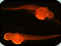
Indeed, zebrafish have emerged as a highly promising and exciting model organism for biomedical research in the life sciences. They offer several advantages over traditional mouse models, making them an attractive choice for many researchers, such as fast reproductive cycle, economical husbandry, optical transparency, easy genetic manipulation, regenerative potential, ethical considerations, and behavioral studies. Zebrafish is also a great tool for imaging due to its transparency. Fully transparent zebrafish, also known as casper zebrafish, are a genetically modified strain of zebrafish that exhibit increased transparency throughout their entire body, including organs and tissues. This transparency allows for enhanced visualization and imaging of internal structures and processes. The development of fully transparent zebrafish has opened up new opportunities for high-resolution imaging and studying complex biological phenomena.
When studying zebrafish, a popular model organism in biology and genetics research, various types of microscopes can be used depending on the specific experimental needs and imaging requirements. Here are some common types of microscopes utilized in zebrafish research:
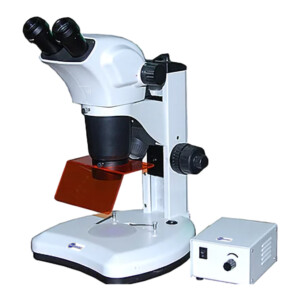
1. StereomicroscopeStereomicroscopes, also known as dissecting microscopes or zoom microscopes, are widely used for examining zebrafish embryos, larvae, and adult fish. They provide low to moderate magnification with a wide field of view, allowing researchers to observe and manipulate zebrafish specimens with ease. Stereomicroscopes are particularly useful for studying morphological characteristics, behavior, and developmental processes in zebrafish. |
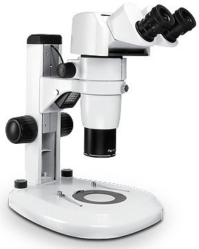 |
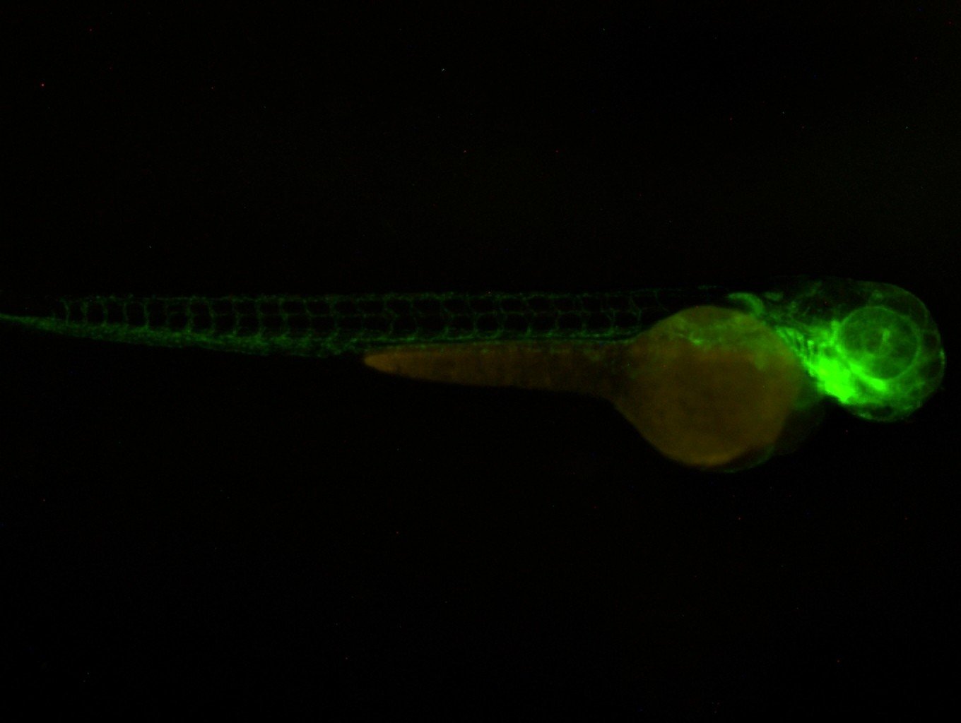
2. Fluorescence Microscope:Fluorescence microscopy is extensively employed in zebrafish research due to the availability of fluorescent reporter genes and fluorophore-labeled probes. Fluorescence stereo microscopes can visualize specific molecules or structures in zebrafish tissues and cells, such as gene expression patterns, protein localization, and cellular processes. This enables researchers to study various aspects of zebrafish development, neurobiology, and disease models. |
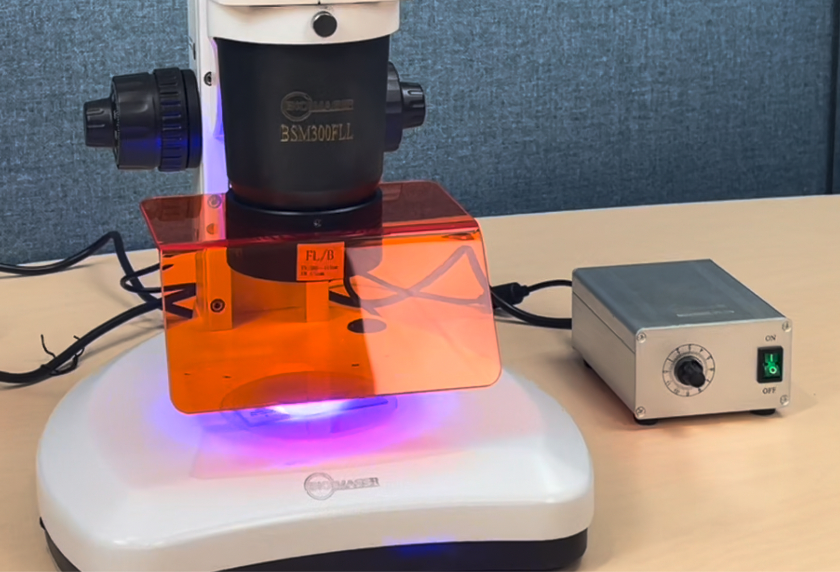 |
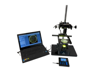
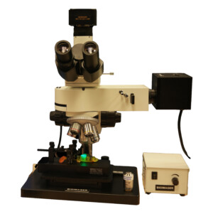
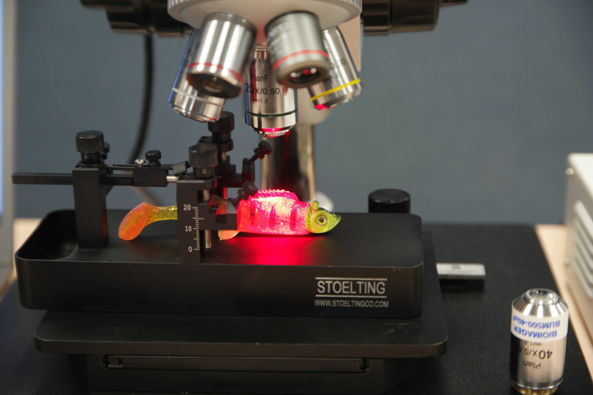
3. In vivo imagingIn vivo imaging of zebrafish refers to the process of visualizing and studying biological processes, tissues, and organs within live zebrafish. This approach allows researchers to observe and analyze dynamic events, developmental processes, disease progression, and drug responses in real-time. Upright or boom stand fluorescence microscopy is widely used for in vivo imaging of zebrafish due to the availability of fluorescent reporter genes and fluorophore-labeled probes. It allows visualization of specific molecules or structures within live zebrafish, such as gene expression patterns, protein localization, and cellular processes. Techniques like confocal microscopy, multiphoton microscopy, or light sheet microscopy can be utilized for high-resolution and 3D imaging of fluorescently labeled zebrafish. |
 |
4. Lightsheet microscopeLight sheet microscopy, also known as selective plane illumination microscopy (SPIM), is well-suited for imaging zebrafish embryos and larvae. This technique uses a thin sheet of light to illuminate the specimen from the side, while a separate objective captures the resulting images. Light sheet microscopy provides high-resolution 3D imaging with reduced phototoxicity and is ideal for long-term, live imaging experiments. |
 |
5. Confocal Microscope:Confocal microscopy is a powerful imaging technique that provides optical sectioning and improved spatial resolution. It is commonly used to visualize specific tissues, organs, or cellular structures in zebrafish with high precision. Confocal microscopes can generate three-dimensional (3D) images and reconstruct zebrafish anatomy or study complex biological processes in detail. |
6. Two-Photon Microscope:Two-photon microscopy is an advanced imaging technique that allows deep imaging of zebrafish tissues without significant photodamage. It utilizes near-infrared light to excite fluorophores, enabling imaging at greater depths within living zebrafish. Two-photon microscopy is particularly useful for studying dynamic processes, neural activity, and cellular interactions in intact zebrafish embryos or larvae. |
7.Electron Microscope:Electron microscopy (EM) offers ultra-high resolution for visualizing fine details in zebrafish tissues and cells. Transmission electron microscopy (TEM) and scanning electron microscopy (SEM) can reveal subcellular structures, cell ultrastructure, and tissue organization in zebrafish samples. EM techniques are commonly used in studies involving zebrafish development, organogenesis, and pathology. |
These are just a few examples of the microscopes used in zebrafish research. The choice of microscope depends on the specific research goals, imaging requirements, and the level of detail needed to answer the scientific questions at hand. If you still need to consult with an imaging expert, feel free to contact us.