Gel & Blot Scanners
Showing all 7 results
-
-
-
-
$USD 18,850.00 Add to cart
-
-
-
Showing all 7 results
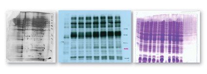
Gel and Western Blots Imaging
A Western blot, also known as an immunoblot, is a laboratory technique used to detect specific proteins in a sample of biological material. The technique involves separating proteins based on their molecular weight using gel electrophoresis and transferring them onto a membrane, which is then probed with a specific antibody that recognizes the target protein.
Here is a general overview of the Western blot process:
- Sample preparation: The biological sample is extracted and treated with a protein denaturant to break down the protein structures.
- Electrophoresis: The protein sample is loaded onto a polyacrylamide gel and subjected to electrophoresis, a process that separates the proteins based on their molecular weight.
- Transfer: The separated proteins are transferred from the gel onto a nitrocellulose or PVDF membrane using an electric current.
- Blocking: The membrane is then blocked with a protein such as BSA or milk to prevent non-specific binding of the detection antibody.
- Primary antibody incubation: The membrane is then incubated with a primary antibody that recognizes the target protein.
- Secondary antibody incubation: A secondary antibody, conjugated to an enzyme or fluorescent tag, is then added to the membrane. This antibody recognizes the primary antibody and binds to it.
- Detection: The enzyme or fluorescent tag on the secondary antibody is then detected using a substrate or imaging system, respectively, which produces a signal that can be visualized and quantified.
Western blotting is a widely used technique in molecular biology and biochemistry, as it allows for the detection and quantification of specific proteins in a complex mixture. It is particularly useful in characterizing protein expression, post-translational modifications, and interactions.
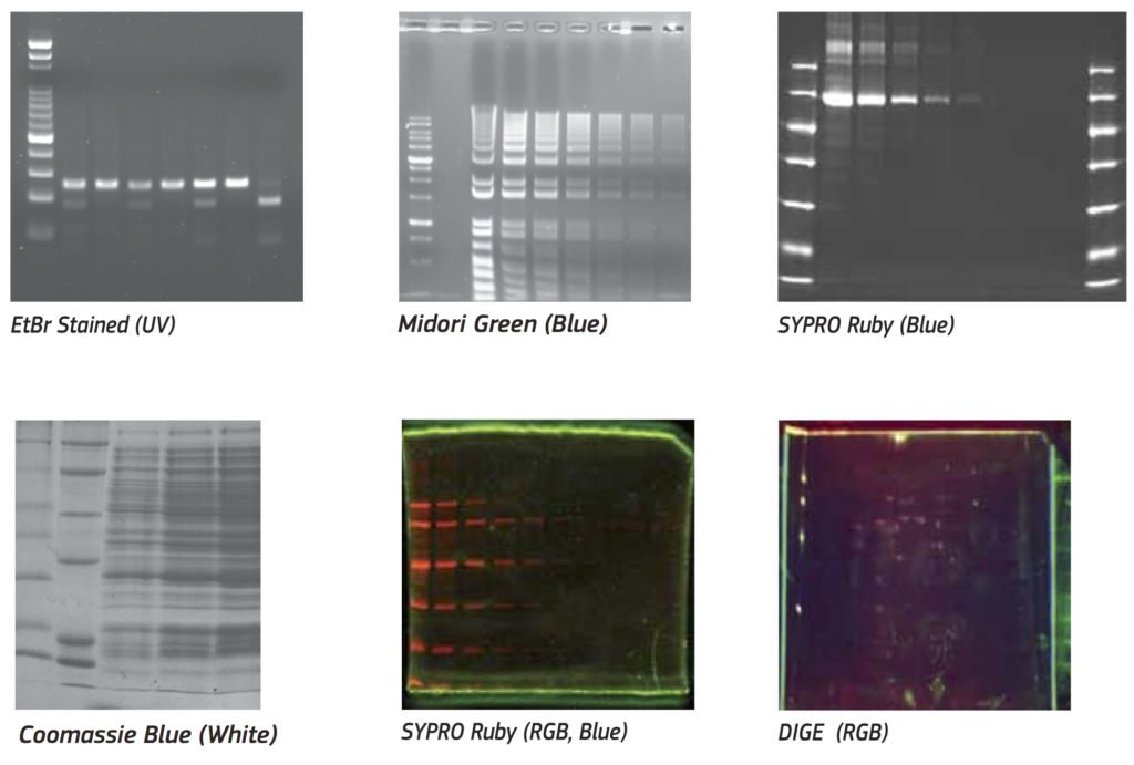
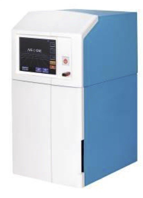
BIOIMAGER’s Gel Imaging Products include Gel Scanner for Electrophoresis Gel, SDS-PAGE, Western Blotting, from low price to professional and advanced models of chemiluminescence gel imaging system. These are more sensitive chemi and colorimetric gel and blot detection in 1D or 2D. You can detect and quantitate multiplex colorimetric, chemiluminescent, fluorescent, and radioisotopic blots.
How to choose a western blot imager?
Choosing a Western blot imager can be a challenging task, as there are many different models and brands available in the market. Here are some factors to consider when selecting a Western blot imager:
- Sensitivity: The sensitivity of the imager is an important consideration, as it determines the level of detection of your protein of interest. The sensitivity of the imager is influenced by the camera, the detection method, and the software used for analysis. Some imagers use chemiluminescence detection, which is highly sensitive, while others use fluorescence detection, which can be less sensitive but allows for multiplexing.
- Imaging system: The type of imaging system used by the imager can also impact its performance. Some imagers use CCD cameras, while others use CMOS cameras. CCD cameras tend to have higher sensitivity and lower noise levels, but they are also more expensive. CMOS cameras, on the other hand, are cheaper but have higher noise levels and lower sensitivity.
- Image resolution: The resolution of the imager is another important consideration, as it determines the level of detail in the images obtained. Higher resolution cameras can produce more detailed images, but they may also have slower acquisition times and higher costs.
- Versatility: The ability of the imager to perform different types of imaging, such as chemiluminescence, fluorescence, and colorimetric detection, is also important. Some imagers can perform multiple types of imaging, while others are more specialized.
- Software: The software used for image analysis is an important consideration, as it determines the accuracy and reproducibility of the results. Look for software that is user-friendly and can perform advanced analysis such as quantification, normalization, and background correction.
- Budget: The cost of the imager is a crucial factor to consider, as it can vary widely depending on the features and capabilities of the instrument. Decide on a budget that meets your needs and look for imagers that offer the best value for money.
Overall, choosing a Western blot imager requires careful consideration of the sensitivity, imaging system, image resolution, versatility, software, and budget. It’s essential to choose an imager that meets your specific requirements and can provide accurate and reliable results.
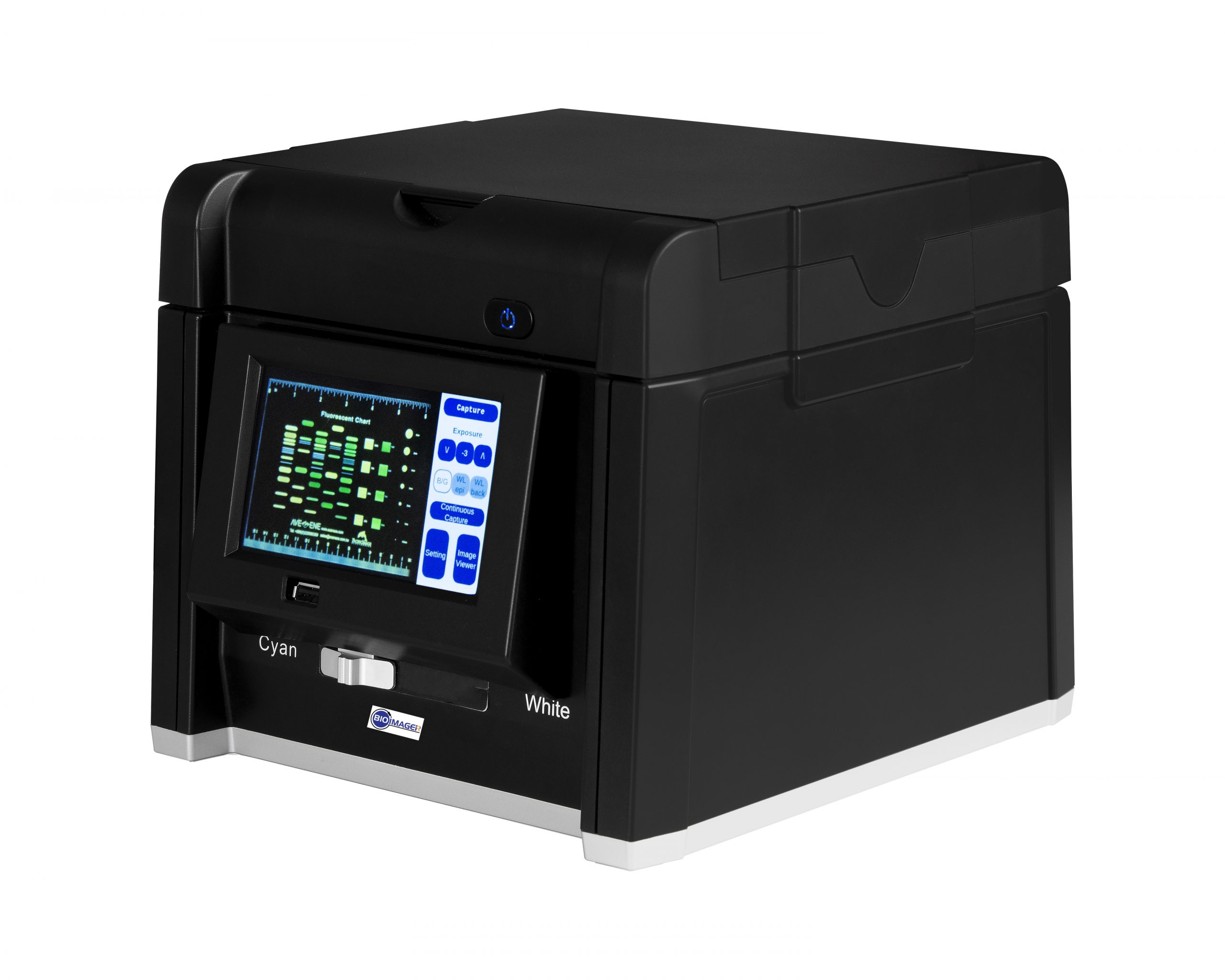
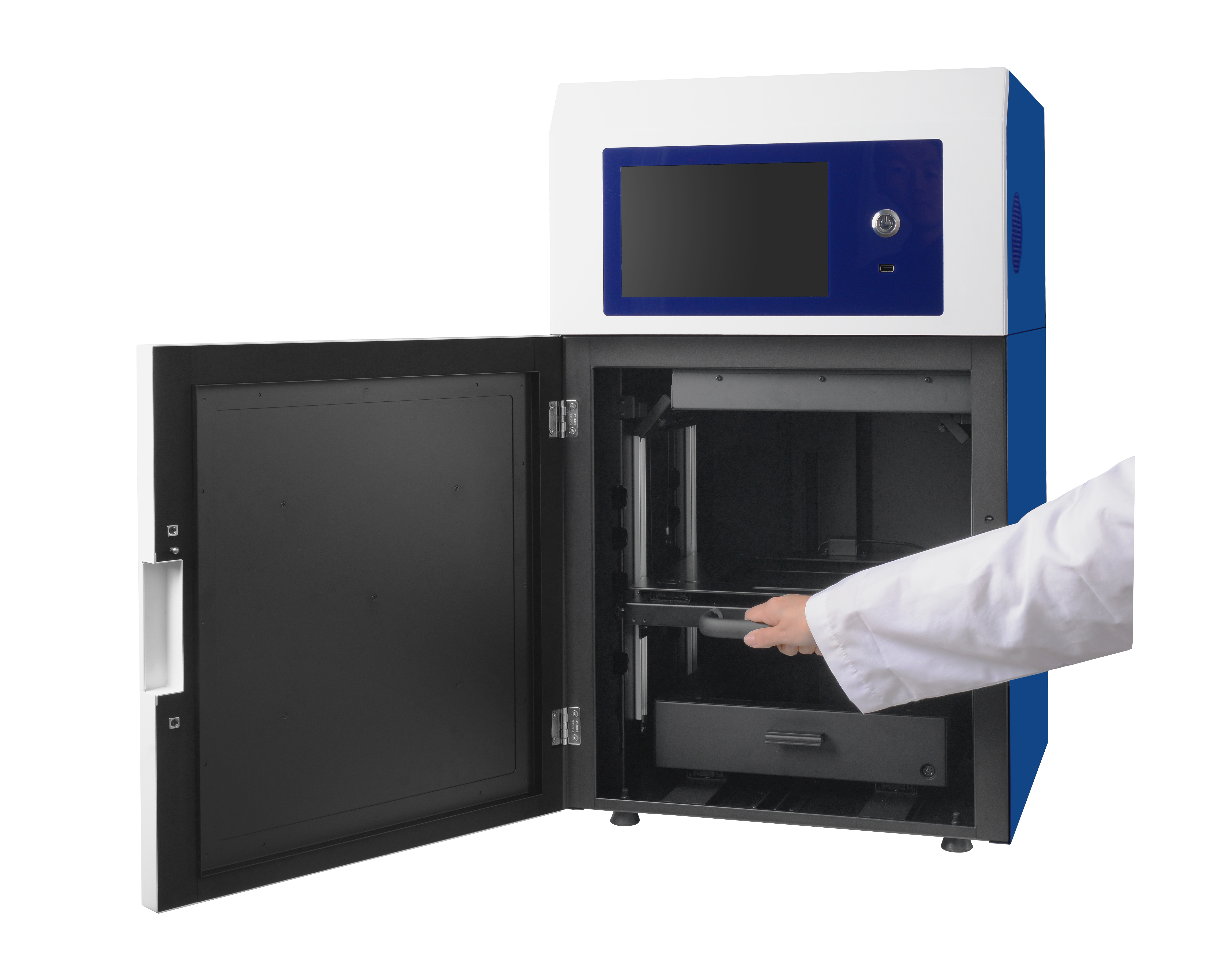
Professional Gel Scanner for Electrophoresis Gel, SDS-PAGE, Western Blotting, etc.
Our Gel Scanner, performing advanced application in biological laboratory, is perfectly designed for image capture of dry or wet sample of electrophoresis gel, SDS-PAGE, western blotting, and so on.
The gel scanner, presenting greater image quality, has great features including super high optical resolution, minimum O.D., which delivers astonishing, sharpness and detailed image for the light area. With the Auto Focus technology, no matter the scanned target is uneven or crease reflective, Microtek’s gel scanner can solve problems, which presents near-original images. Besides, only Bioimager provides the Emulsion Direct Imaging Technology (E.D.I.T.), which efficiently eliminates problems such as Newton rings and surface imperfection. The Microtek’s gel scanner is the best choice to digitize gel images and offer a total solution for biological professionals and researchers.
Thanks to the design of gel scanner, the glass holder can be removed anytime. The glass holders can be cleaned and washed easily. Scientists can capture images directly from gel surface handily no matter the samples are dry or wet. It saves a lot of time, 1-2 days at least, to continue the lab projects without waiting sample dry.
To get more information of this smart gel scanner with excellent image quality, please contact our Sales team.
What is Chemiluminescence Imaging?
Chemiluminescence is the process of light emission resulting from a chemical reaction. When certain chemicals react, they can produce excited-state molecules that release energy in the form of light. This process is similar to bioluminescence, where organisms such as fireflies and certain marine animals produce light through a chemical reaction.
Chemiluminescence is commonly used in analytical chemistry and biochemistry for the detection of substances that are not easily detected by other means. For example, in forensic science, chemiluminescence is used to detect trace amounts of blood at a crime scene.
Some examples of chemiluminescent reactions include the reaction of luminol with hydrogen peroxide in the presence of a catalyst such as iron or copper, which produces a blue light, and the reaction of fireflies with oxygen and luciferin, which produces a yellow-green light.
Why to choose Chemiluminescence?
Chemiluminescence is used for various purposes in different fields due to its unique properties. Here are some common uses of chemiluminescence:
- Analytical Chemistry: In analytical chemistry, chemiluminescence is used as a detection method for various compounds, including proteins, DNA, and other biomolecules. The process of chemiluminescence is highly sensitive, allowing the detection of low levels of analytes.
- Biochemistry: In biochemistry, chemiluminescence is used for studying enzyme reactions, protein-protein interactions, and for detecting reactive oxygen species (ROS) and free radicals. It is also used in immunoassays to detect specific antibodies and antigens.
- Medical Diagnostics: In medical diagnostics, chemiluminescence is used for detecting specific biomarkers and proteins in blood, urine, and other bodily fluids. This technology is used to diagnose diseases such as cancer, infectious diseases, and autoimmune disorders.
- Forensic Science: In forensic science, chemiluminescence is used to detect trace amounts of blood, semen, and other bodily fluids at a crime scene. This method is highly sensitive and can detect small amounts of biological material that may not be visible to the naked eye.
- Environmental Monitoring: In environmental monitoring, chemiluminescence is used to measure the levels of pollutants such as ozone, nitrogen oxides, and sulfur dioxide in the atmosphere. The technology is also used to monitor the quality of water and soil.
Overall, chemiluminescence is a powerful tool that offers high sensitivity, specificity, and versatility in various fields, making it a valuable analytical and diagnostic method.