Metallurgical Microscopes
Showing 1–9 of 71 results
-
-
-
 Request a Quote Request a Quote
 -
-
-
-
-
-
 Request a Quote Request a Quote
Showing 1–9 of 71 results
Metallurgical Microscopes
Introduction
Metallurgical microscopes, also known as materials microscopes, come in different configurations such as upright, inverted, or boom stand, to accommodate various sample sizes. An upright metallurgical microscope is used for smaller samples that fit on the microscope stage, while an inverted metallurgical microscope is designed for larger parts, allowing heavy mechanical components to be placed directly on the stage above the objectives. A boom stand metallurgical microscope provides a greater working distance than an upright microscope when needed.
While metallurgical microscopes may resemble compound biological microscopes, they possess distinct characteristics. They enable high-magnification viewing of samples (up to 500x and 1000x) without light passing through the sample, as in biological microscopy. Although stereo microscopes offer reflected illumination, metallurgical microscopes surpass them in resolution and magnification.
Metallurgical microscopes offer several features that set them apart from biological or stereo microscopes, including polarization, differential interference contrast (DIC), high-power magnification, high-resolution reflected light illumination, and options for brightfield and darkfield imaging. They utilize reflected illumination and sometimes transmitted illumination as well.
Metallurgical microscopes find application in examining various samples, such as metal parts, fiberglass, industrial manufacturing failures, carbon fiber, plastics, and concrete.
When choosing a metallurgical microscope, consider the illumination type—reflected or reflected and transmitted—and the objective lens options, as some microscopes support only brightfield microscopy while others offer darkfield capabilities. The size of the samples also plays a role in determining the appropriate microscope configuration. Smaller samples suit an upright metallurgical microscope, while larger samples like car parts benefit from an inverted setup. If you have any questions regarding the ideal metallurgical microscope for your needs, feel free to contact Bioimager.
Applications
Metallurgical microscopes are used for a variety of applications, including inspection of electronic components, measurement of precision machined parts, and examination of materials for defects and structural analysis. They are often equipped with specialized imaging systems, such as high-resolution cameras, polarizers, and differential interference contrast (DIC) optics, that allow for detailed examination and analysis of metallurgical materials and components.
Metallurgical /Industrial microscopes are also often equipped with advanced software that allows for real-time imaging and analysis of the samples. This software can be used for measurement and analysis of features such as particle size, shape, and distribution, and can also be used for defect detection and classification.
Overall, metallurgical microscopes are a valuable tool for industrial applications and manufacturing, and they play a critical role in ensuring product quality and reliability.
A reliable microscope is an essence for effective diagnosis and professional measurements. The microscopist performs the inspection of materials, prepare an analysis report then a supervisor makes a proper decision. There are so many details on the objects that cannot be seen by a regular microscope. Suitable microscopy parts, optomechanical components, camera, and software are required to achieve such a goal.
BIOIMAGER’s metallurgical microscopes offer high magnification with reflected and transmitted light. Metallograph microscopes allow the user to view opaque items at high magnification. Specialty uses for metallurgical microscopes include use as measuring instruments for measuring thin films, electroplating coatings, grain size, surface inclusions and defects. Our metallurgical microscopes are available in binocular and trinocular models with both upright, inverted and boom stand microscope systems.
Metallurgical microscopes are available with brightfield only or brightfield and darkfield. There is a great explanation of imaging modes here and metallurgical microscopy basics and application here. The high magnification of metallurgical microscopes is perfect for viewing items that won’t allow light to pass through them (such as metals, coated films and solid objects where defects must be detected.) All metallurgical microscopes have the option to add a microscopy camera and software for capturing images and making measurements.
1. Metallurgical Microscope Types
Metallurgical microscopes are divided mainly to 4 categories:
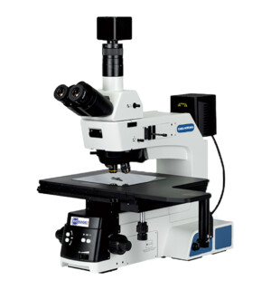
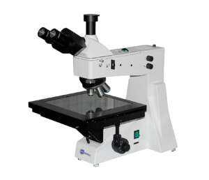
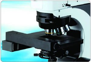
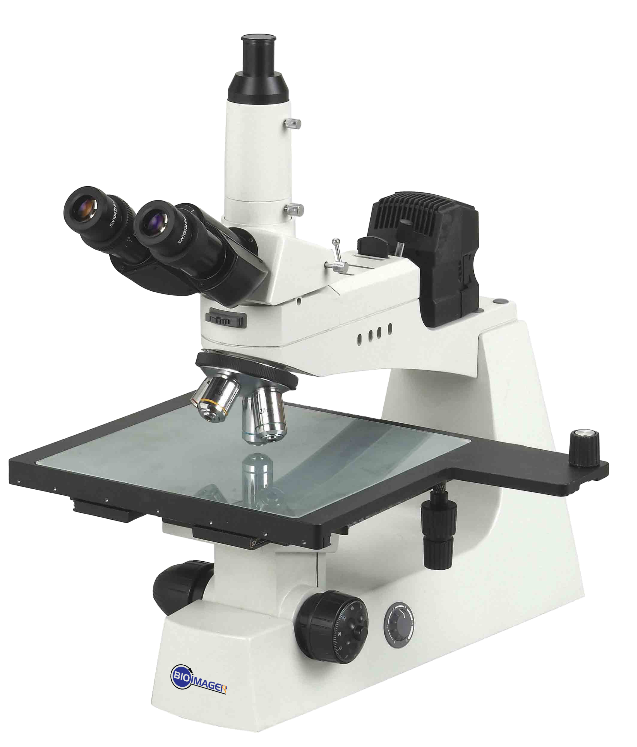
1) Upright Microscopes with reflected light only, in which the light comes from top lamp-house and is used for non-transparent samples
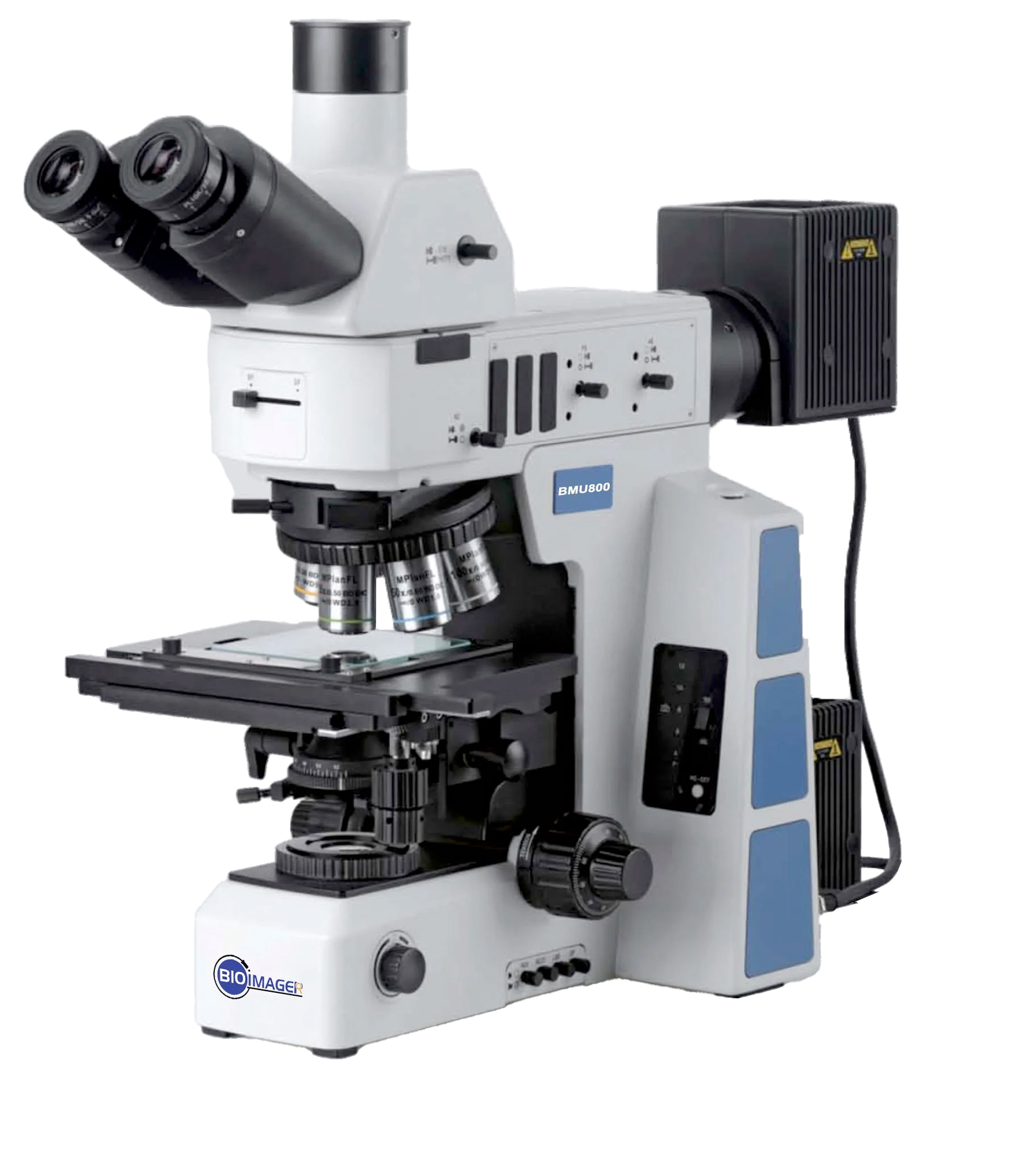
2) Upright Metallurgical Microscopes with reflected and transmitted lights, in which light can come from top and bottom light sources and can be used to examine the transparent and non-transparent samples.
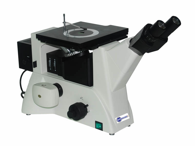
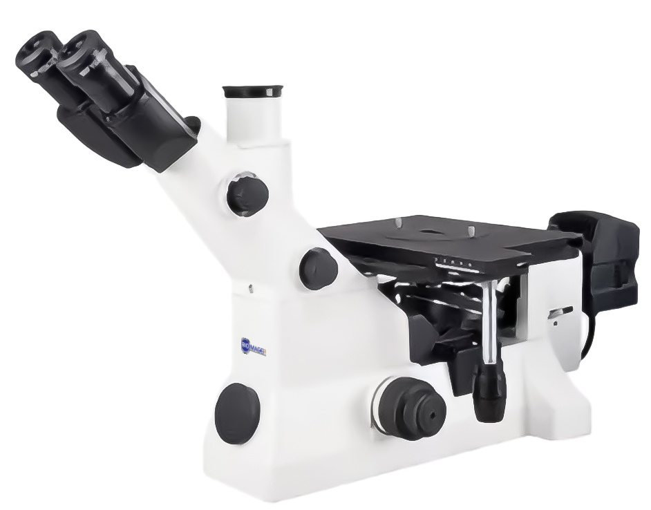
3) Inverted metallurgical Microscopes which includes a lamp house for reflected illumination. Bioimager offers economic external light source to be able to use for transparent samples
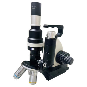
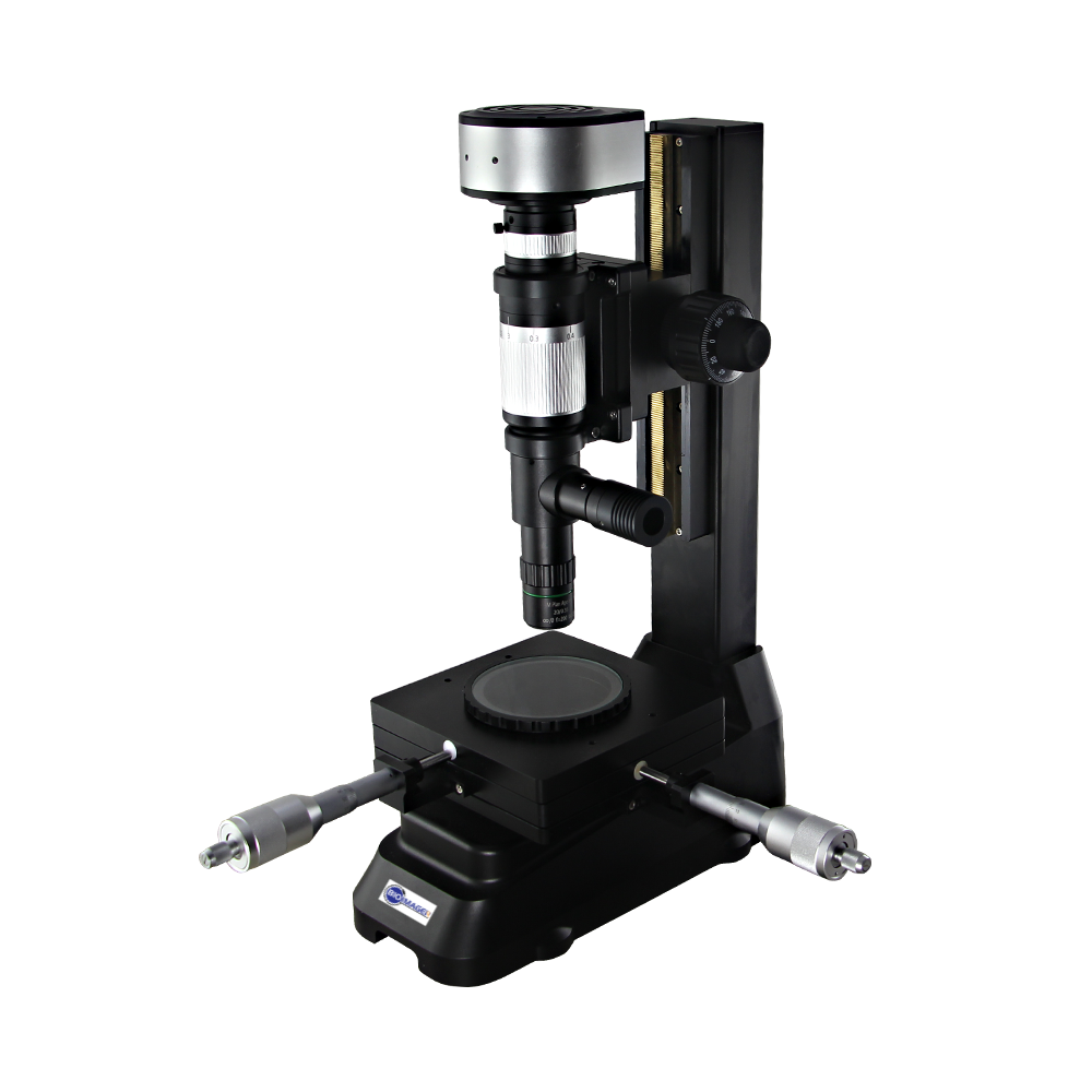
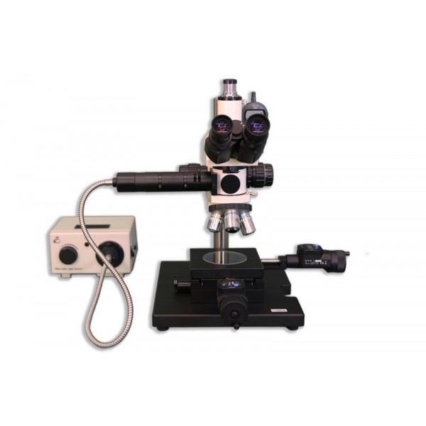
4) Boom Stand Metallurgical Microscopes
 |
 |
 |
 |
| Upright Metallurgical Microscope with Reflected light only | Upright Metallurgical Microscope with Reflected & Transmitted | Inverted Metallurgical Microscopes | Boom Stand Metallurgical Microscopes |
Please select the products from the left side category list or search bar.
2. Imaging Capabilities
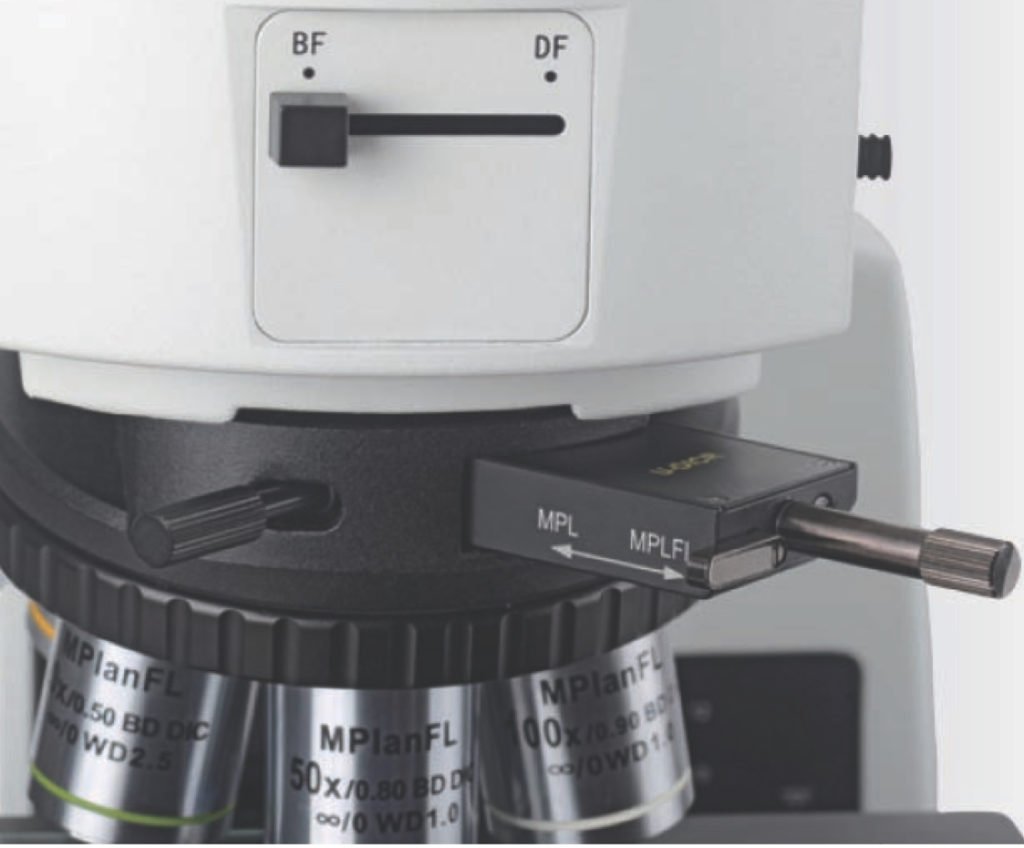
a) BF or DF? By default all metallurgical microscopes come with brightfield (BF) imaging. If you need to have darkfield (DF) imaging, you do need to decide during the purchase time as upgrading such a feature will not only be costly but also will result in loosing the money you spent on BF module and also the obj lenses. Switching between BF and DF is always super easy, just move a lever.
b) DIC Nomarsky or not? DIC Nomarsky imaging needs a polarizer and analyzer. Make sure your microscope does / will come with polarizer, analyzer, slot for DIC insert and also which objective lenses are supported for DIC imaging.
Having DIC Nomarsky is always costly. If your microscope allows having a DIC imaging but your budget does not permit, make sure you can upgrade that by simply purchasing the extra parts without discarding your current obj lenses or setup.
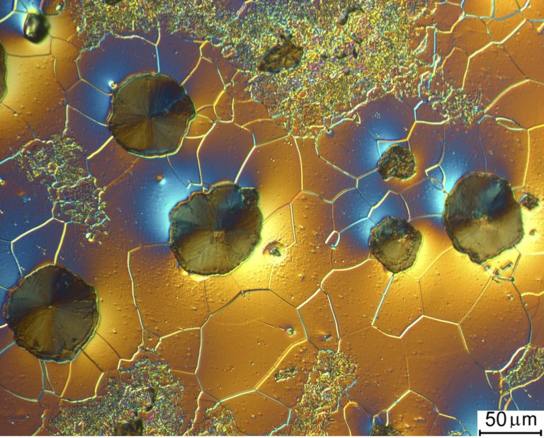
Differential-Interference-contrast image of cast iron with spheroidal graphite, 500X magnification, captured with ProgRes® C14
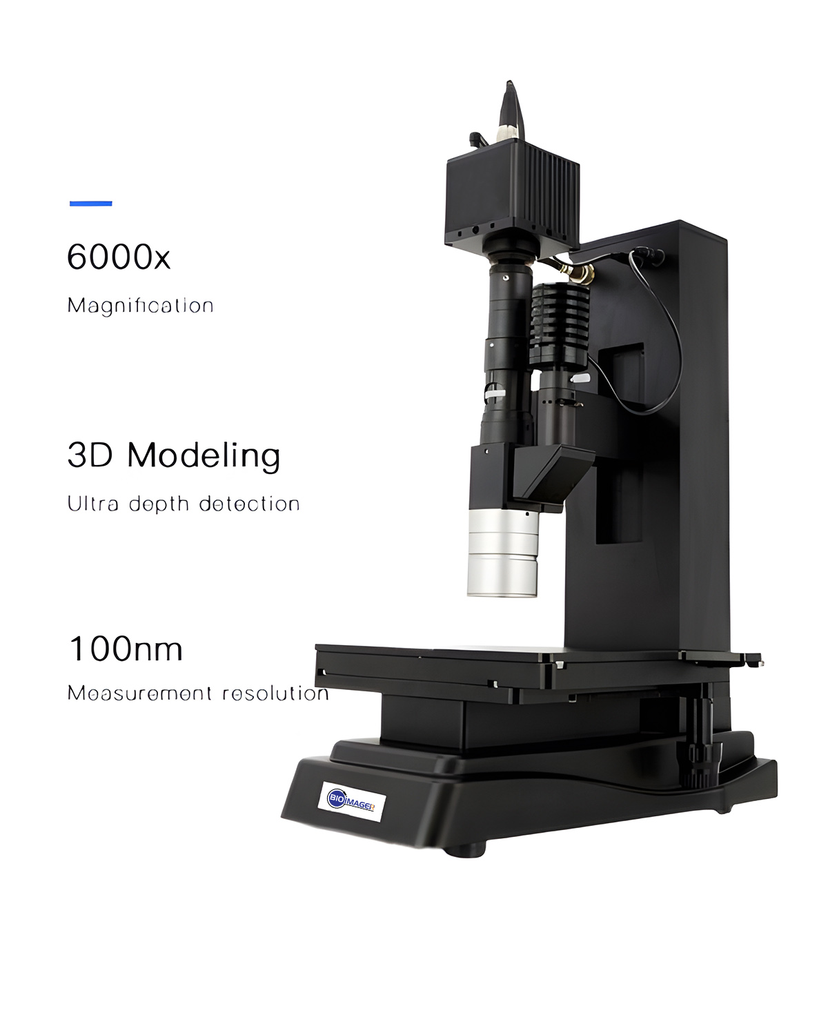
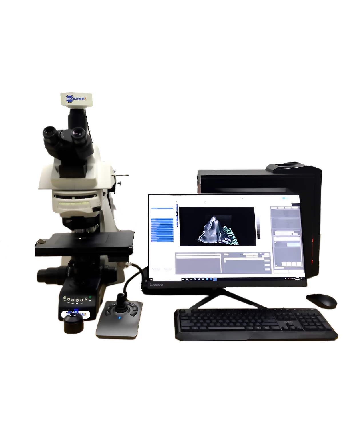
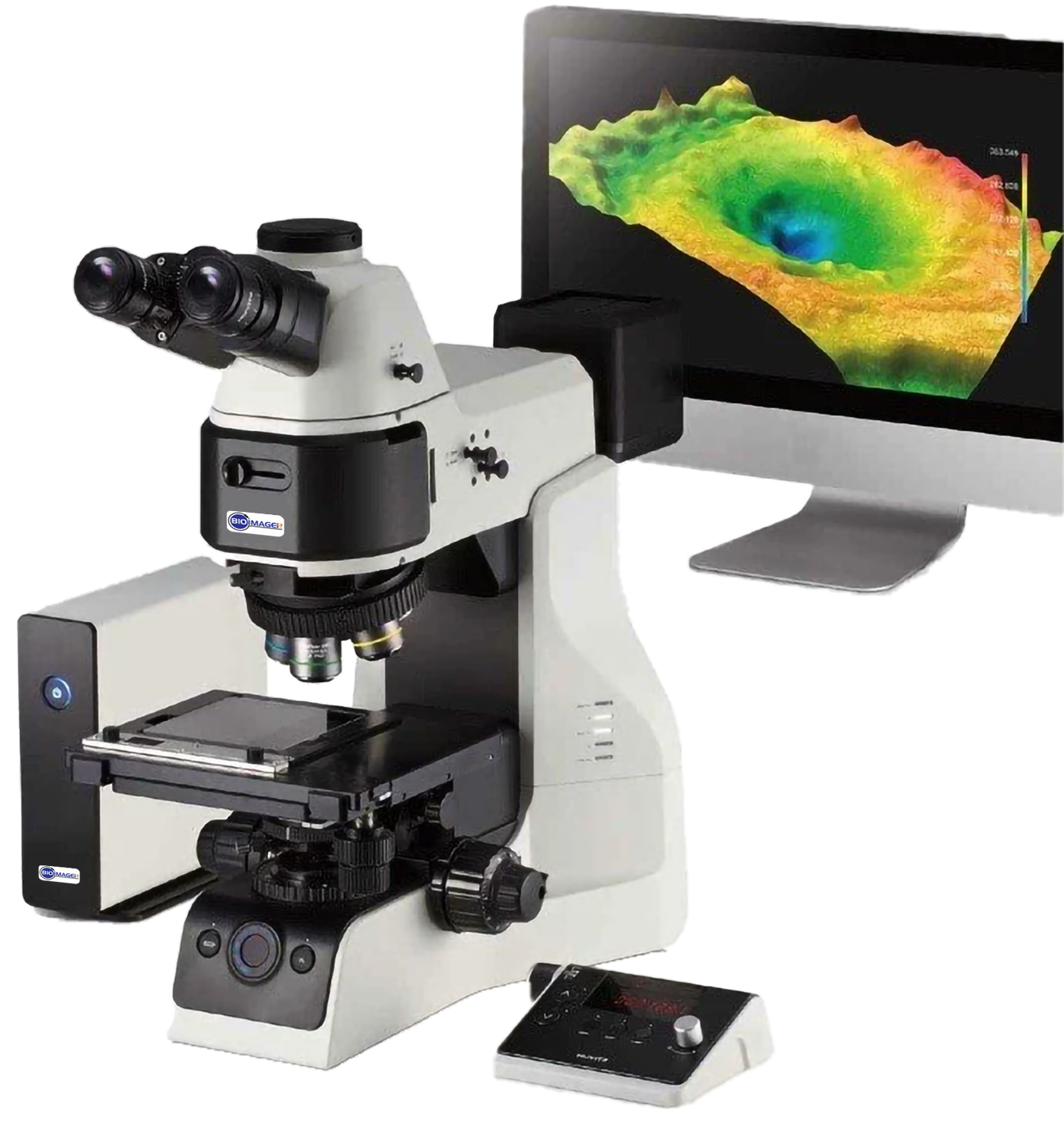
3. The 3D Profiler
one of the most interesting and useful feature of the metallurgical microscopes is having a 3D profiler which allows measurement of the height and/or thickness of a sample. This is a great tool for inspection of the materials or scaffold are made in three-dimensional, such as 3D tissue or dental cements. BIOIMAGER has recently added an output format of .stp to the constructed 3D microscopy images then you can easily print out what you observe under microscope.
 |
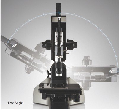 |
| 3D Profile with automated Z focus at 10 nm resolution | 3D Profiler with tilting stand |
4. Illumination Guide for Transmitted & Reflected Light
Transmitted Light
| Specimen Type | Imaging Technique |
| Transparent Specimens – Bacteria, spermatozoa, cells in glass containers, protozoa, mites, fibers, etc. | Phase Contrast
Oblique Illumination |
| Light Scattering Objects – Diatoms, fibers, hair, fresh water microorganisms, radiolarians, etc | Darkfield Illumination
Phase Contrast |
| Light Reflecting Specimens –
Colloidal suspensions, powders and minerals, liquids |
Phase Contrast
Dispersion Staining |
| Amplitude Specimens –
Stained Tissue, naturally colored specimens, hair & fibers, insects and marine algae |
Brightfield Illumination |
| Fluorescent Specimens –
Cells in tissue culture, fluorochrome-stained sections, smears and spreads |
Fluorescence Illumination |
| Birefringent Specimens –
Mineral thin sections, liquid crystals, melted and re-crystallized chemicals, hairs & fibers, bones & feathers |
Polarized Illumination |
Reflected Light
| Specimen Type | Imaging Technique |
| Specular (Reflecting) Surfaces –
Thin film, mirrors, polished metallurgical samples, integrated circuits |
Brightfield Illumination
Darkfield Illumination |
| Diffuse (Non-Reflecting) Surfaces – Thick and thin films, rocks & minerals, hair, fiber, bone and insects | Brightfield Illumination
Darkfield Illumination Polarized Illumination |
| Amplitude Surface Features –
Dyed fibers, diffuse metallic specimens, composite materials, polymers |
Brightfield Illumination
Darkfield Illumination |
| Birefringent Specimens – Mineral thin sections, hair & fibers, bones & feathers, single crystals, oriented films | Polarized Illumination |
| Fluorescent Specimens –
Mounted cells, fluorochrome-stained sections, smears and spreads |
Fluorescence Illumination |
5. Analysis and Measurements
| Field / Sample | Image | Attributes, Measurement Features, Analysis |
| Ceramics, composites, Plastic |  |
|
| Food |  |
|
| Metals |  |
|
| Particles & Porous materials |  |
|
| Cosmetics | 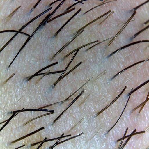 |
|
| Glass, films, coatings | 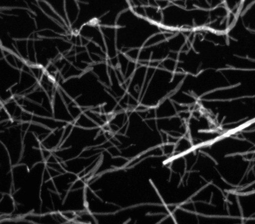 |
|
| Textile / Fibers | 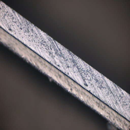 |
|
| new materials | 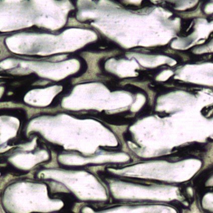 |
|
These features can be analyzed with Image Pro Plus or Image Pro Premier.
Analysis Example: Fiber Separation Analysis
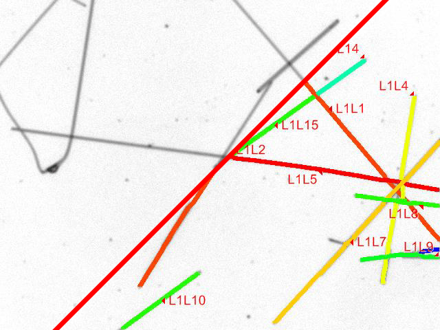 |
The application is designed to separate and measure crossing fibers on images. The fibers on the images can be straight or bent. It can be used to measure distribution of thin inclusions or asbestos fibers.
Researchers have been looking for an easier way to measure the lengths and widths of overlapping fibers. Most researchers end up manually measuring fibers – a process that is not only time consuming, but can introduce inconsistencies. Now, with the Image-Pro Premier Fiber Separation App, it is possible to measure and classify the length and thickness of overlapping fibers with one click. This App is designed for applications where the entire fiber is visible in the image. |
6. Sample Images / Applications
 |
||
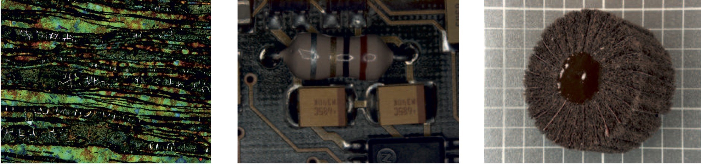
| Sample: Duplex steel, damaged rotor blade etching: Beraha III, Camera: ProgRes® C5, Courtesy of: Cloeren Technology/Germany | Sample: PC board, Camera: ProgRes® C14, Courtesy of: ProgRes® Application Lab | Sample: Blower grinding tool, Camera: ProgRes® C14plus, Courtesy of: ProgRes® Application Lab |
 |
||
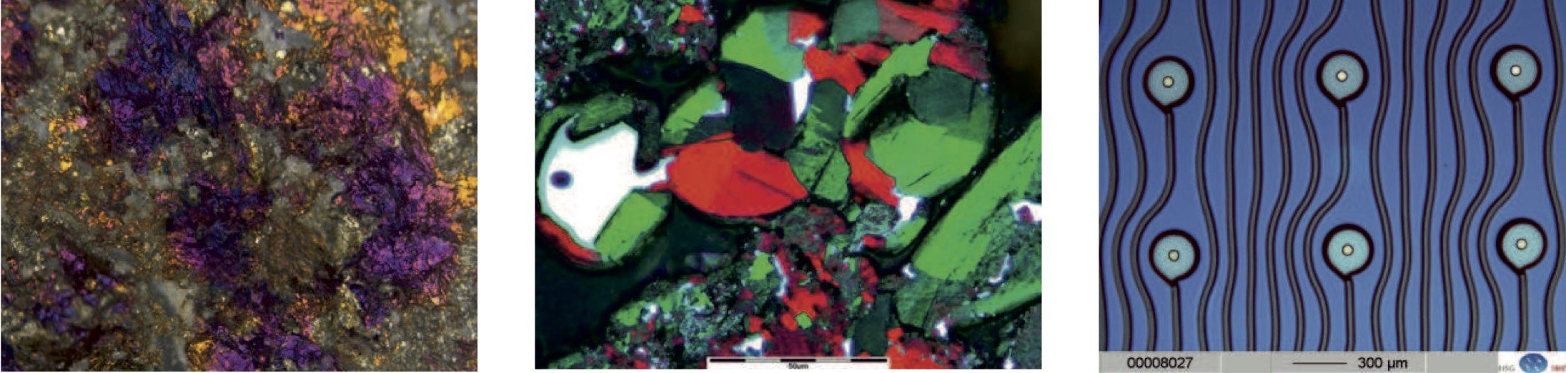
| Sample: stone on stereo microscopes Camera: ProgRes® C7, Courtesy of: ProgRes® Application Lab | Sample: compound from graphite and powder, Camera: ProgRes® C3, Courtesy of: Hoffmann & Co. Elektrokohle AG, Austria | Sample: fluid channel fo a press button Camera: ProgRes® 3012, Courtesy of: HSG-IMIT Microtechnology |
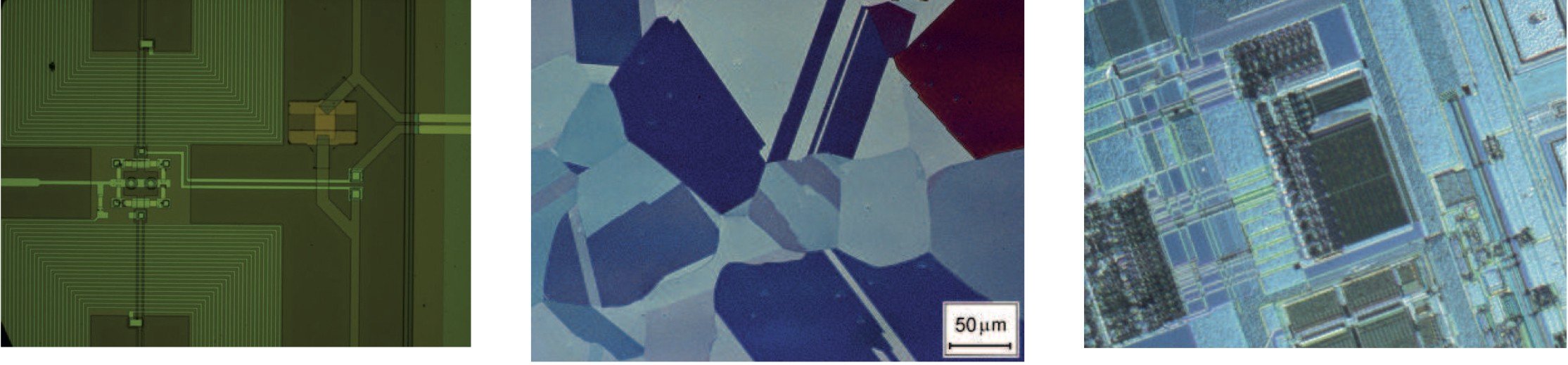 |
||
| Sample: detail of a SQUID (superconducting quantum interference device), Camera: ProgRes® C3, Courtesy of: MPQ of Radio Astronomy, Bonn, Germany | Sample: rolled metal sheet, Camera: ProgRes® C14, Courtesy of: MPI for Iron Research GmbH | Sample: Wafer im DIC Camera: ProgRes® C14plus Courtesy of: Promicron |
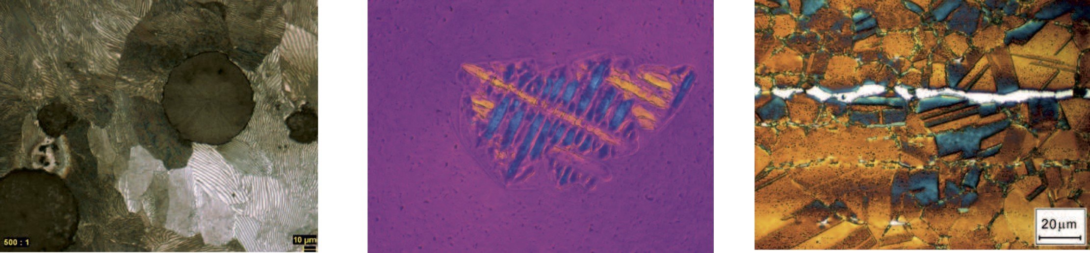 |
||
| Sample: Cast Iron (spherulitic graphite iron), Camera: ProgRes® C5, Courtesy of: Cloearen Technology, Germany | Sample: new formation of cristobalite in glass, Camera: ProgRes® C14plus, Courtesy of: Institute of Ceramic, Glass and Building Materials of the Mining Academy | Sample: TWIP Steel, Camera: ProgRes® C14, Courtesy of: MPI for Iron Research GmbH |
7. Video Tutorial: How To Use a Compound Light Microscope
See Polarized and DIC Imaging Video tutorials at their pages.