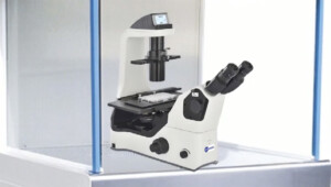
BUM400AFL is an inexpensive imaging package designed specifically for scanning of a single or double glass slide in brightfield and epi-fluorescence mode with optional darkfield, phase-contrast and polarizing imaging modes. Reflected imaging mode is also available on request.
Why BUM400AFL? This vs competitor products in the market.
There are two popular whole slide-scanning microscopes in the market. Both are made by two brands of Zeiss and Leica. Their price is at 6 digits. Ours are way below that and you will save at least 50% in the price. We understand our microscope is not as fast as those products, but its quality of images remains reasonably great. On top of that we offer this at small packages, which is affordable for small businesses and new PIs in the universities. Our winning point is that lots of customization can be offered such as custom filter sets per your application, camera resolution and sensitivity per your selection and range of objective lenses.
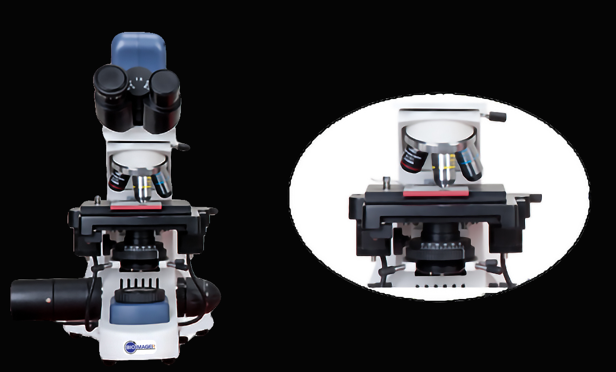
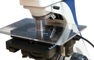
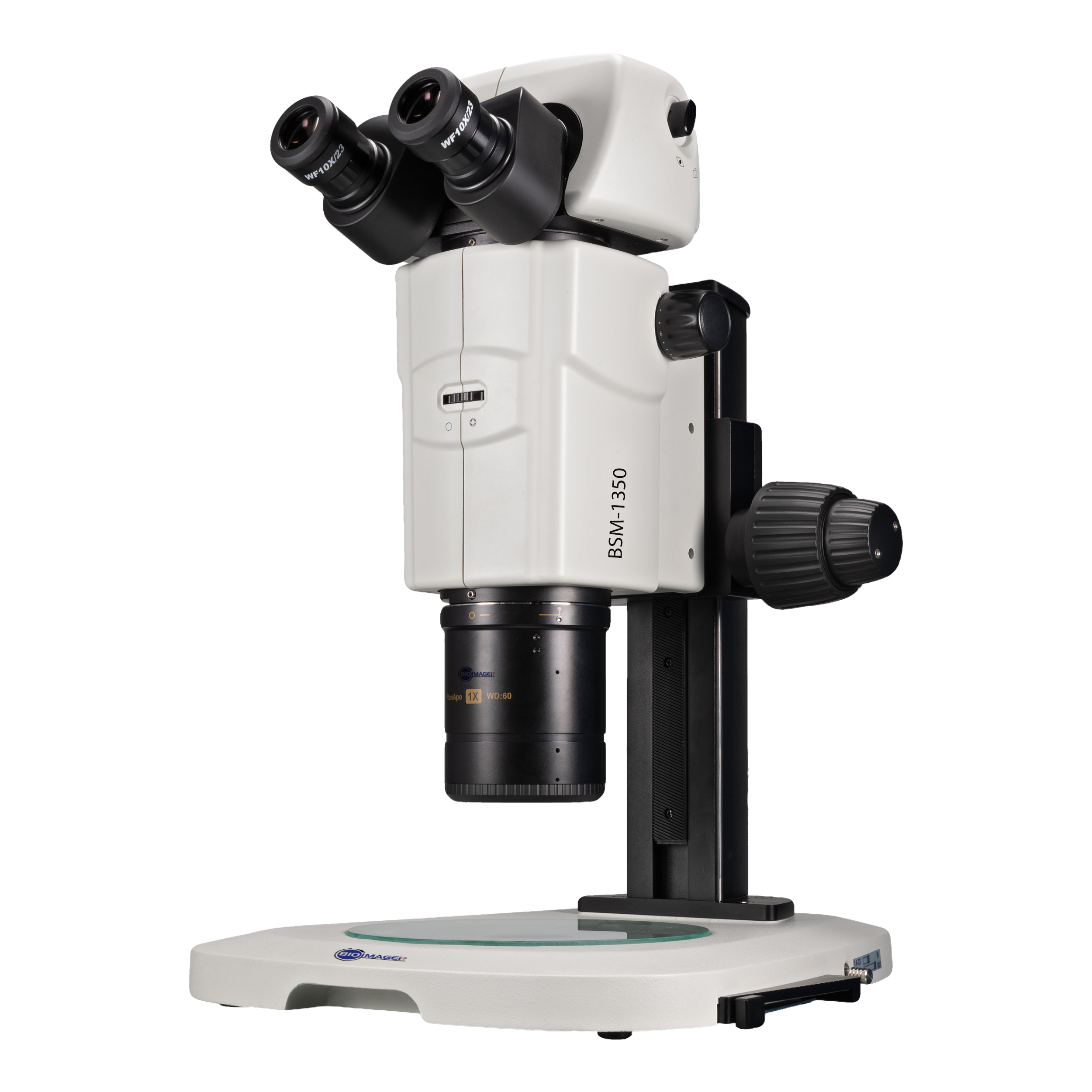
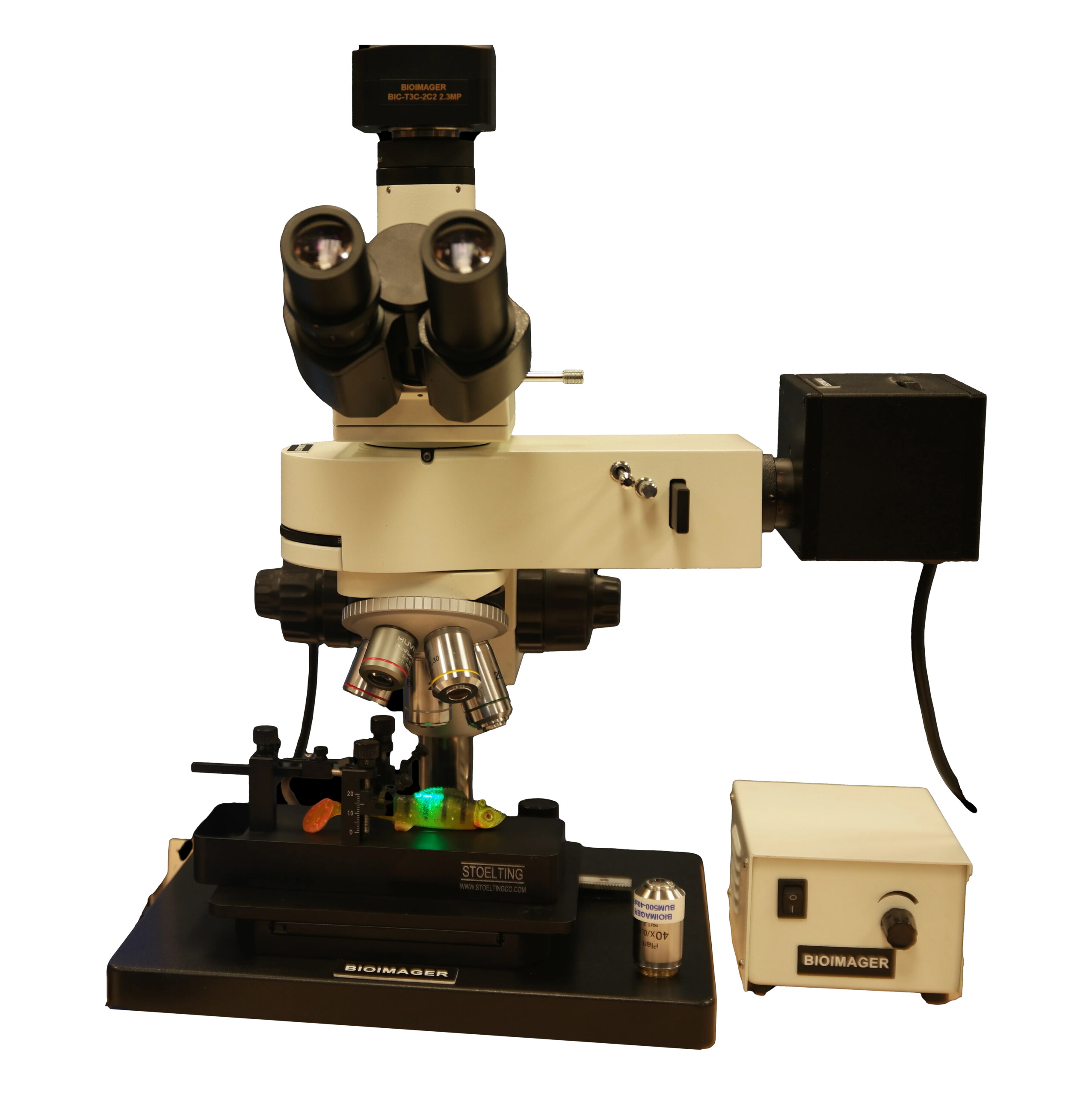
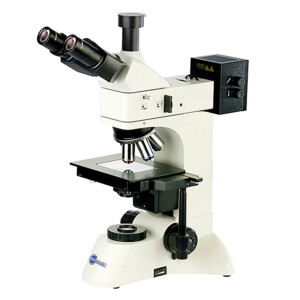
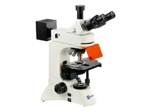
The BUM400AFL is an automated upright biological microscope with LED fluorescence imaging specifically designed for scanning of fluorescent glass slides or any fluorescent samples can be visualized from top view.
BUM400AFL is our recent product designed specifically for scanning of a single or double glass slide in brightfield and epi-fluorescence imaging with optional darkfield and polarized imaging. Reflected mode is also available upon request.
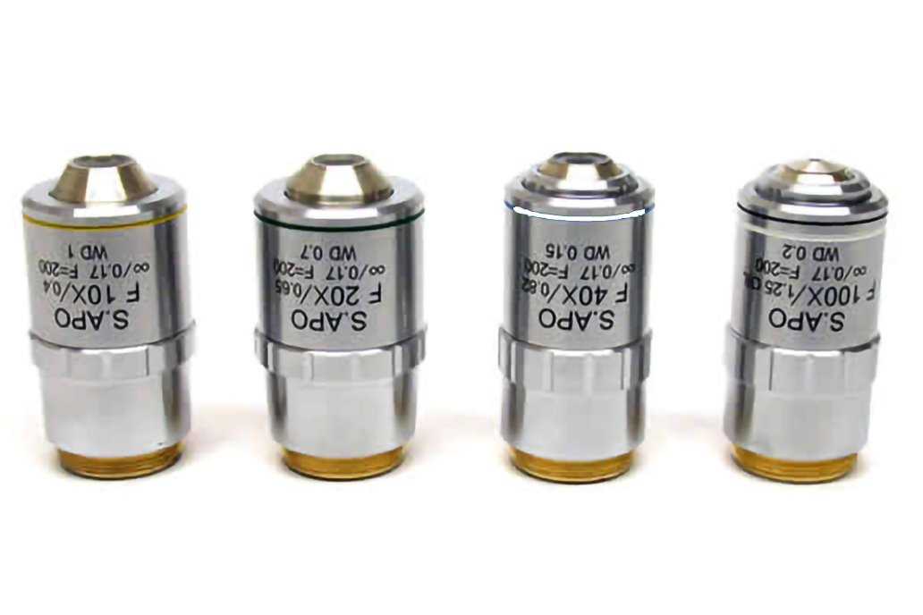
The system includes by default 4x, 10x, and 20x objective lenses but it can be upgraded easily or selected from the beginning with other lenses. Low magnification lenses are great for quick scan of the whole slide view. For this purpose, we have 1.25x, 2x, 2.5x, 4x and 5x objective lenses. It happens the customer struggles deciding between 20x and 40x objective lenses, as 20x has less resolution but 40x requires at least four times more scanning time of 20x lens. For this type of application, we suggest using 25x objective lens, which is at middle point between 10x and 40x. If you need a high magnification over 40x, you can select 50x (dry or oil), 60x (dry or oil), or 100x (oil).
Whole Slide Imaging
For whole slide imaging, a 2x obj lens does a great job. It takes 3-4minutes to scan whole slide. If you use a 1.25x obj lens this will be even way better. It takes just 1-2 minutes to complete the full scan.
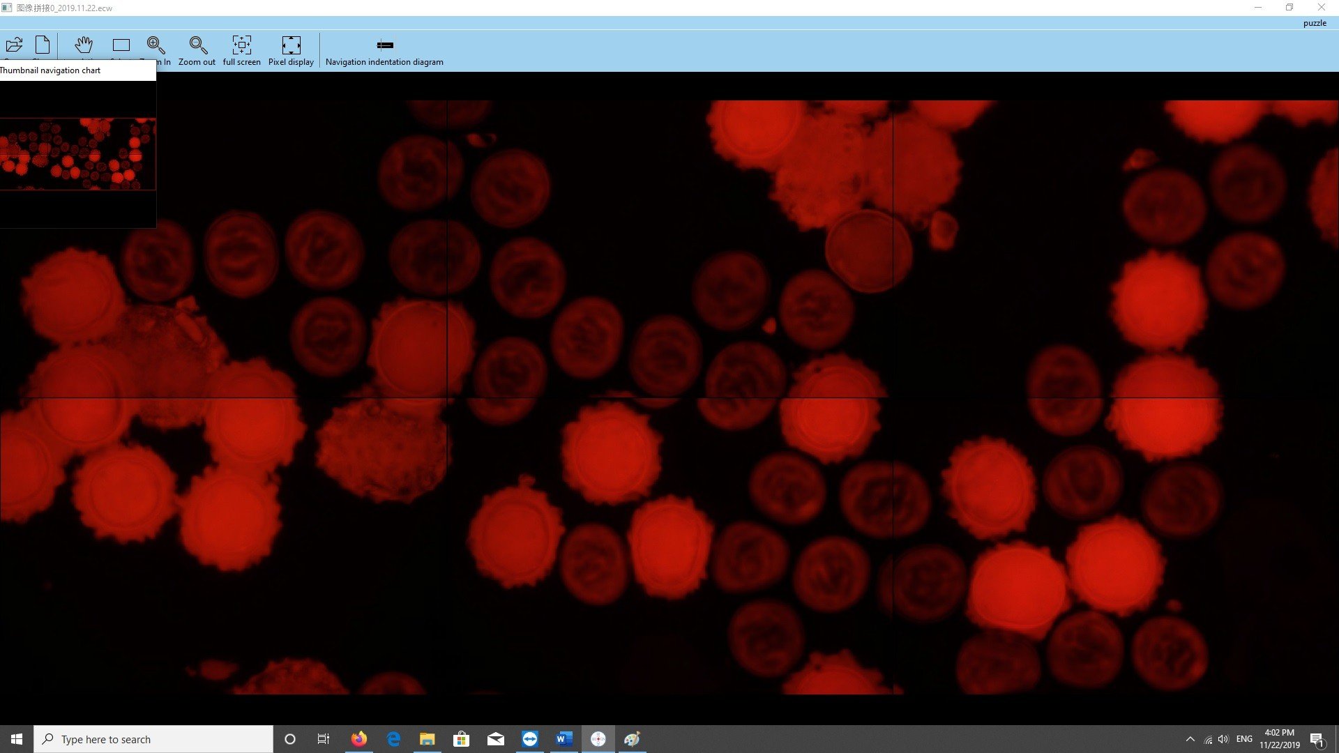
A fluorescence slide for mercury/HBO light alignment with green fibers inside.
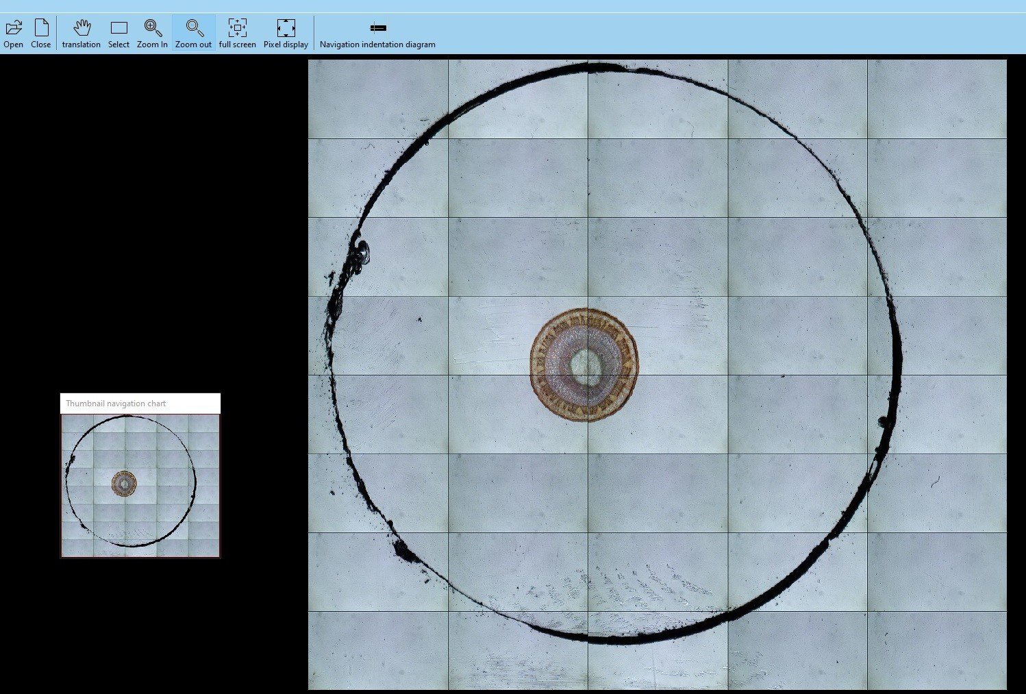
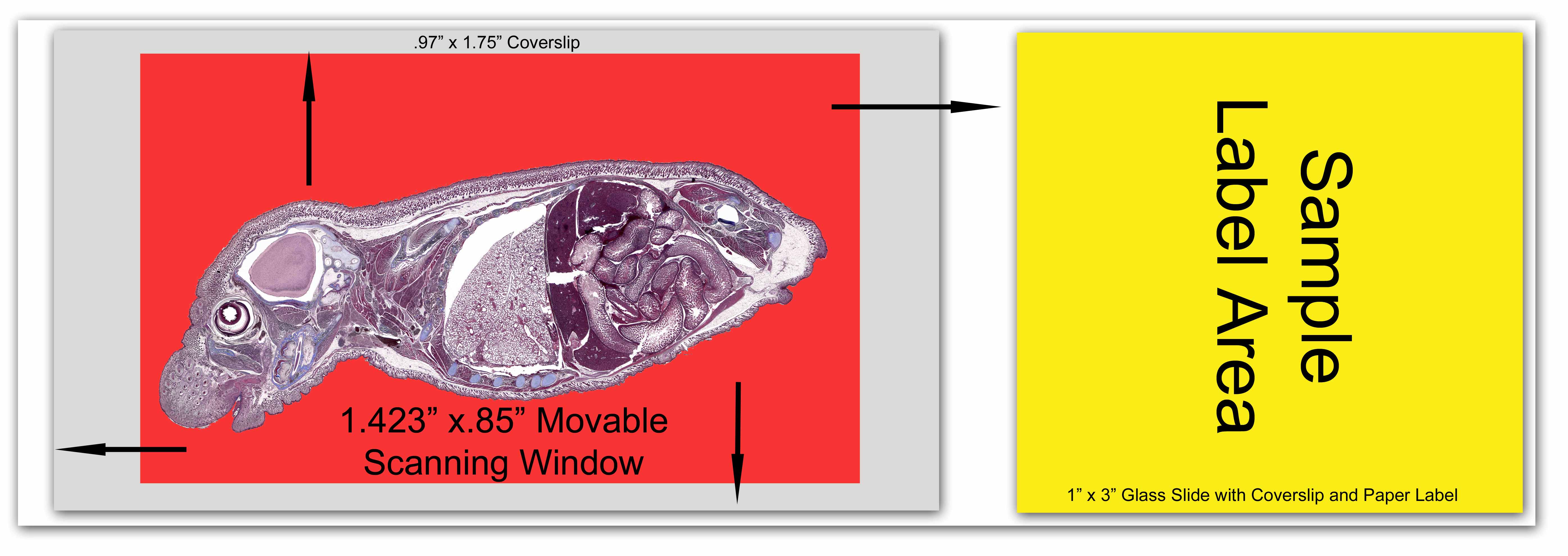
Scanning area with BUM400AFL can be the full size of the glass slide or just the coverslip.
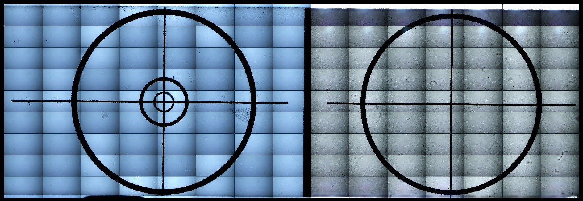
A 20x22mm area is scanned by 2.5x obj lens with a 5×8 tile and processed in less than 94 seconds (1min 34 sec). Below is the raw image with tiled boarder lines.
Please check the “imaging” tab for further sample images.
Field of Views
This table shows the field of view size (Length x height) in micrometers x micrometers using a 2.0MP (1920×1080) Camera and a 0.65x C-Mount adapter:
Objective Lens
Field of View
Pixel Size (um)
2.0x
5,000 um x 2,813 um
2.604
2.5x
4,400 um x 2,475 um
2.292
4x
2,438 um x 1,371 um
1.270
10x
988 um x 556 um
0.515
20x
510 um x 287 um
0.266
40x
233 um x 131 um
0.121
Speed
Time for image acquisition and image processing (stitching the tiles together and displaying) depends on the sample size and imaging condition. The number of tiles or mosaics depends on the sample size and what objective lens is selected, as well as the individual field of view (FOV) size. An FOV size itself is determined by the camera sensor sensor size and the c-mount adapter magnification.
Imaging condition includes the type of imaging (brightfield, fluorescence, darkfield, etc), light intensity, exposure time (and gain determined by the camera type) and most importantly using autofocus condition. Auto-focusing is not required at low magnifications such as 2x, 2.5x, 4x or 5x. If you use a thin sample, this may not be still required at 10x obj lens. For 10x, you may choose partial autofocusing, for instance every 3, 5 or 10 spots of view. Depending on the sample type and if a high magnification such as 20x or above is selected, you may need to do the autofocuisng at each FOV. Meanwhile, the focus range and the number of steps for autofocusing , as well as speed of focusing is matter.
Prior running a big experiment, it is highly recommended to spend enough time to find the right setting parameters for acquisition such as exposure time and focusing parameters. Run small size of experiments, watch the result, tune the parameters then increase the whole size of experiments.
Here is a list of examples to get an idea of the speed using BUM400AFL.
Objective Lens
Spot Size
Tile Size
Imaging Condition
Spent Time(Acquisition Time + processing Time = Total (min: sec)
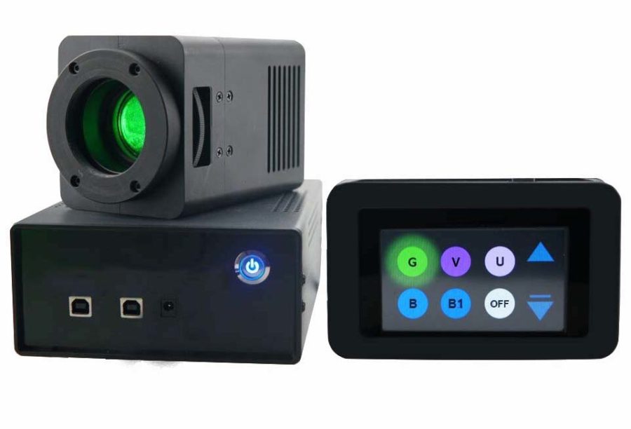
The microscope use a 3W LED for UV, blue and green channels. Custom LEDs and 5W power are also available.
Technical Specification
1. Microscope Optics
DESCRIPTION
SPECIFICATION
Included
Optical System
Basic Unit
BUM400A Automated Microscope
1
Control System
BUM400A-CS for XY stage and Z Focus
1
Eyepiece tube
30°hinged, binocular, (55mm-75mm)
1
Objectives
Plan 4x, 10x, 20x, 40x
1
Revolving nosepiece
Can hold up to 4 objectives
1
Eyepieces
WF 10×/ 22
2
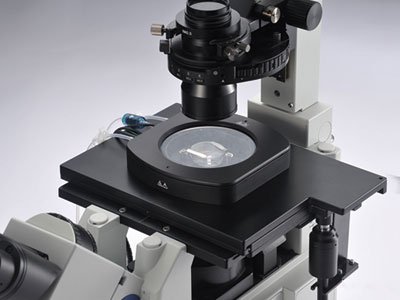
Condenser
N.A 1.25 Abbe condenser with variable diaphragm and filter
1
Power Adapter
110V or 220 V
1
Focus
Fine/Coarse coaxial focus to use gear rack transmission mechanism with 0.002 mm resolution
1
Filter
1
Lamp
High brightness Kohler illumination, Adjustable Halogen lamp 12V/20W (or its equal brightness LED)
AC 85V – 230V
1
Serial/USB data cable
BUM400A Automated Microscope exclusive
1
Stage
Travel range: 50x75mm
Speed: 1 to 50 mm/sec
1
Computer
Intel Core i5 or higher / 500 GB HDD/DVD / 8GB
with 19’’ LED
Optional
Others
Slide Loader
automatic slide loader for racks of 100, 200 slides
Optional
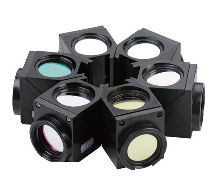
2. Fluorescence Module
LED*
Fluorophores
Filters
Name
EX
Name
EX
EM
EX
DM
EM
Ultraviolet (UV)
365nm
Dapi / Hoechst
350 nm
470 nm
330-385nm
400
420-470nm
Blue light (B)
470nm
FITC / GFP
490 nm
525 nm
460nm-495nm
505
510-550
Green light (G)
505nm
Alexa Fluor 555
555 nm
580 nm
460nm-550nm
570
590-650
Red (R)
(Optional)
625nm
Alexafluor 647
650 nm
665 nm
600-660nm
660
660nm
Custom
400-1000nm
visible, IR
* The specs of LED and filters may change per the customer request.
** Ask us to customize the LEDs and Filters among our over 40 filter sets.
We also have 5W LED source. Please check this product BIA-FL-IS5.
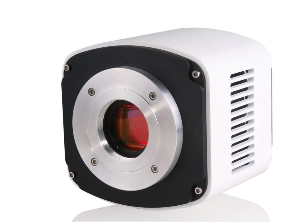
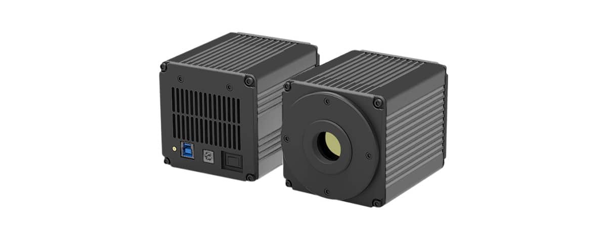
3. Camera
a) CMOS Cameras
Order Code
Sensor &
Size (mm)
Pixel (μm)
G Sensitivity
Dark Signal
FPS/Resolution
Binning
Exposure
BIC-E3S-2.0
2.0MP
IMX385(C)
1/2″(7.20×4.05)
3.75 x3.75
2350mv with 1/30s
0.15mv with 1/30s
125 @1920×1080
1×1
0.1ms ~15s
BIC-E3S-3.1G
3.1MP
IMX265 (C, GS)
1/1.8″(7.07×5.30)
3.45×3.45
1146mv with 1/30s
0.15mv with 1/30s
53 @2048×1536
85 @1024×768
1×1
0.1ms ~15s
BIC-E3S-5G
5.0MP
IMX264(C, GS)
2/3” (8.45×7.07)
3.45×3.45
1146mv with 1/30s
0.15mv with 1/30s
35 @2448×2048
50 @1224×1024
1×1
0.1ms ~15s
BIC-E3S-9.0G
9M
IMX305 (C, GS)
1″(14.13×7.45)
3.45 x3.45
1146mv with 1/30s
0.15mv with 1/30s
34@4096×2160
60@2048×1080
1×1,
2×2,
0.1ms ~15s
BIC-E3S-20.0
20MPIMX183(C)
1″
(13.06×8.76)
2.4 x 2.4
462mv with 1/30s
0.21mv with 1/30s
15@5440×3648
50 @2736×1824
60@1824×1216
1×1,
2×2,
3×3
0.1ms~15s
Note: C: Color, M: Monochrome, GS: Global Shutter
b) CCD, please visit this link: https://www.bioimager.com/product-category/cameras/ccd-cameras/2-stage-cooling/
c) sCMOS, Please visit this link: https://www.bioimager.com/product-category/cameras/scmos/
Videos & Images:
A) Videos
Video 1: Quick software review and fluorecsnce slide imaging
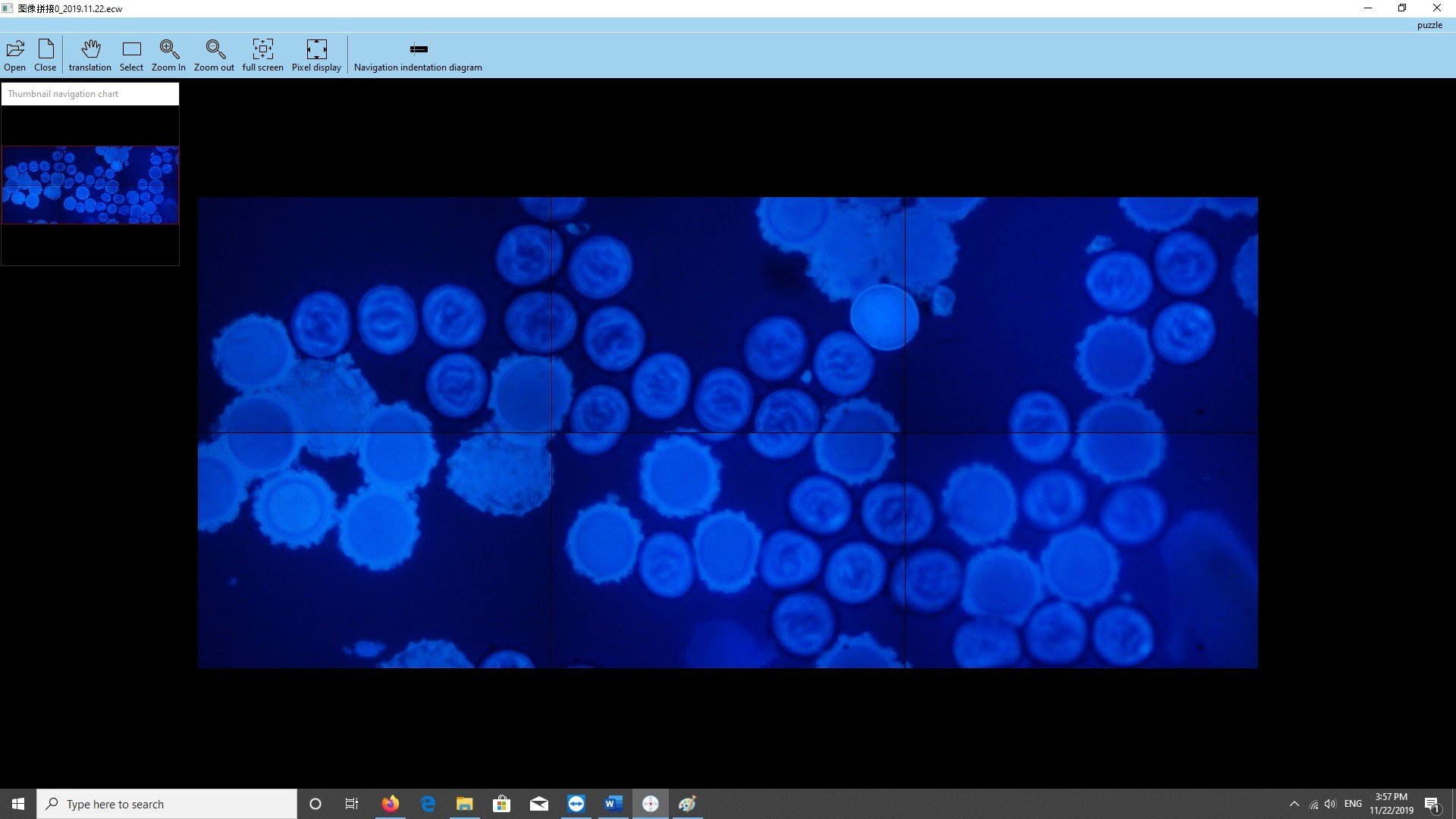
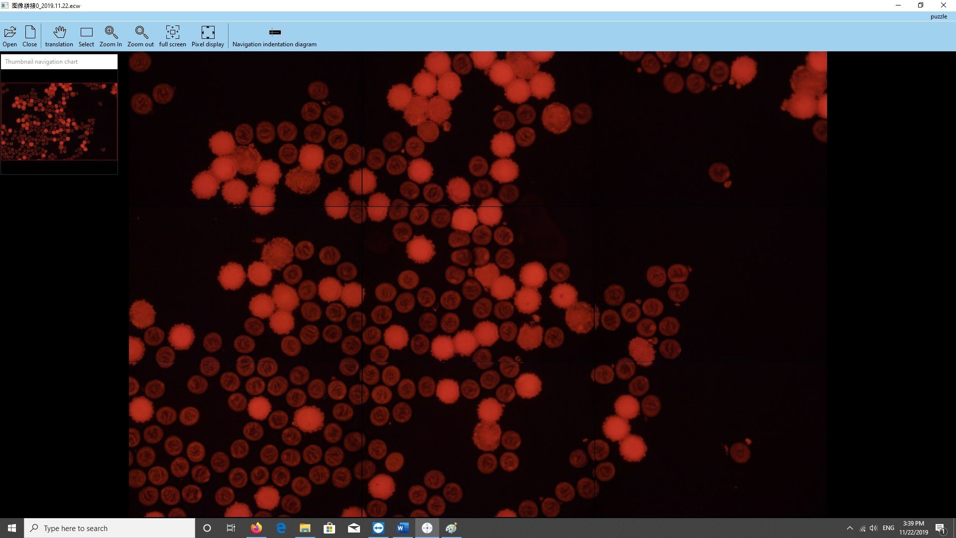
This video demonstrates an example of fluorescence (in red channel) imaging of a potato thin-section slide using 20x objective lens. It overview the software setting for this case study, shows autofocusing, manual exposure and also reviews the final image after stitching 18×21 tiles.
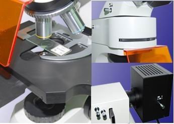
Video 2. Upgrade to reflected brightfield and darkfield imaging with a custom closed miniature incubator
Video 3. Phase Contrast Kit and Sample Images (optional imaging capability)
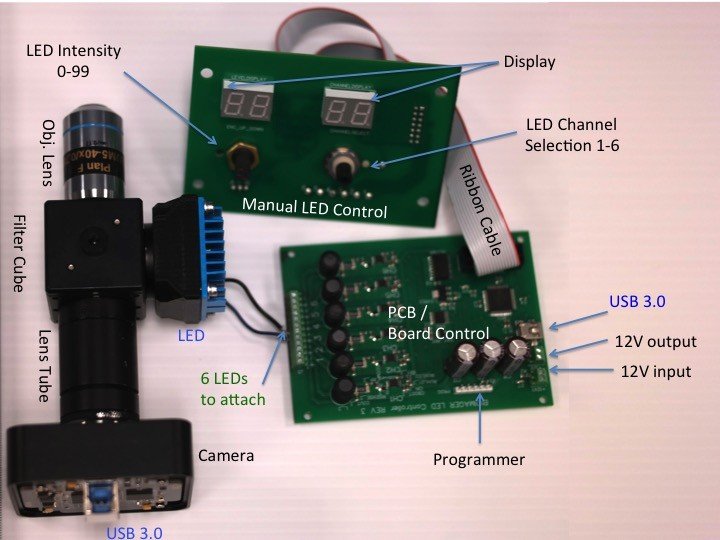
BUM-PHK Phase Contrast Kit for Upright Biological Microscopes is composed of 10x, 20x, 40x , 60x and 100x (oil) Phase contrast objective lenses, green filter, Phase alignment tool and turret condenser with different annulus.
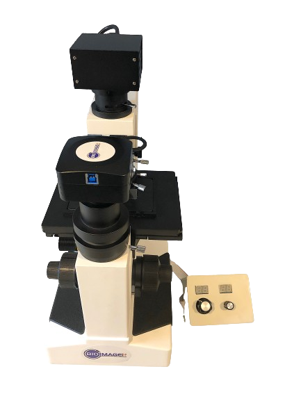
In this video we look at the samples images of an Onion Skin thins section slide and Tilia (basswood) thin section slide, using this kit installed on Bioimager BUM800FL upright biological microscope and connected a 2.3MP USB3.0 CMOS Colorful Camera as well as a 0.5x C-Mount Adapter.
cope and connected a 2.3MP USB3.0 CMOS Colorful Camera as well as a 0.5x C-Mount Adapter.
B) Images
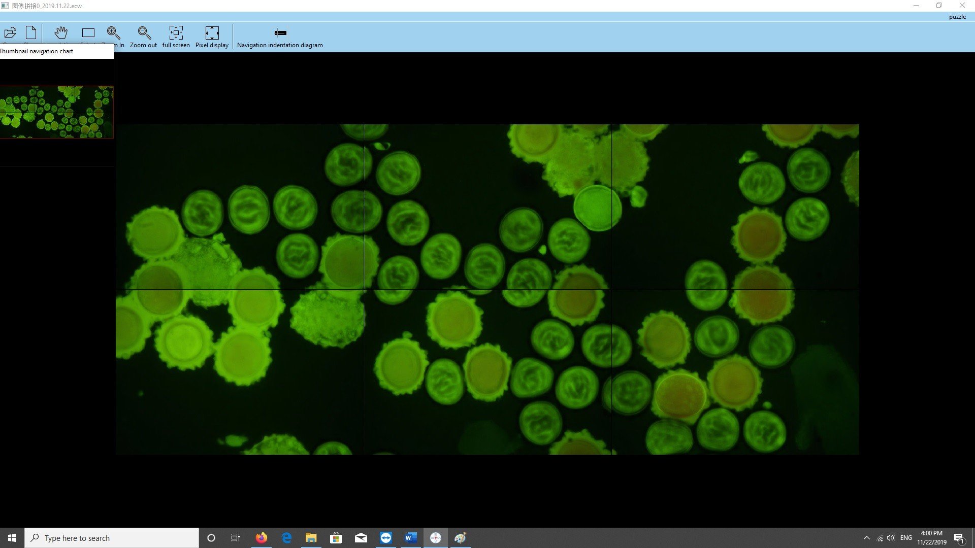
Whole Coverslip 20mm x 20mm Imaging
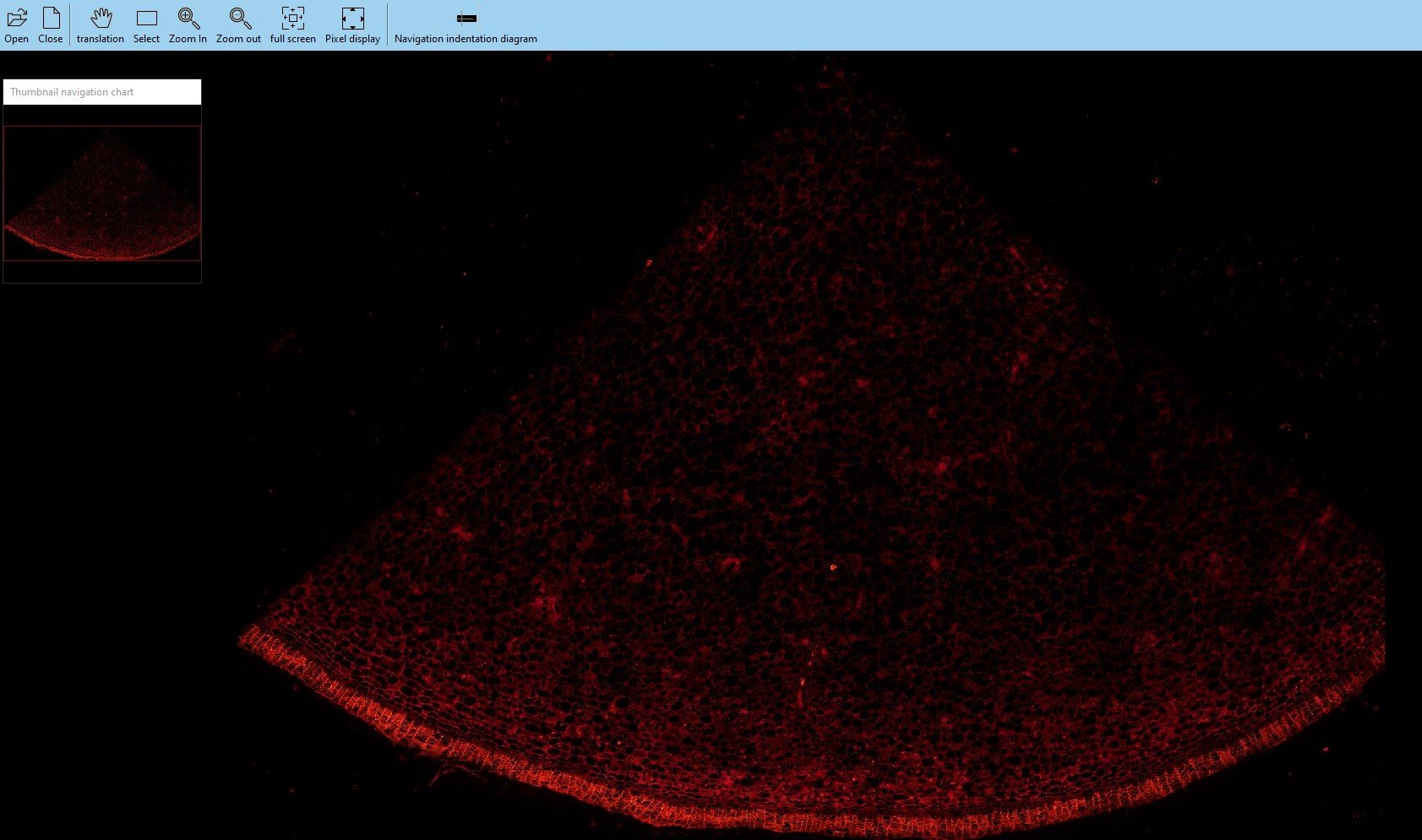
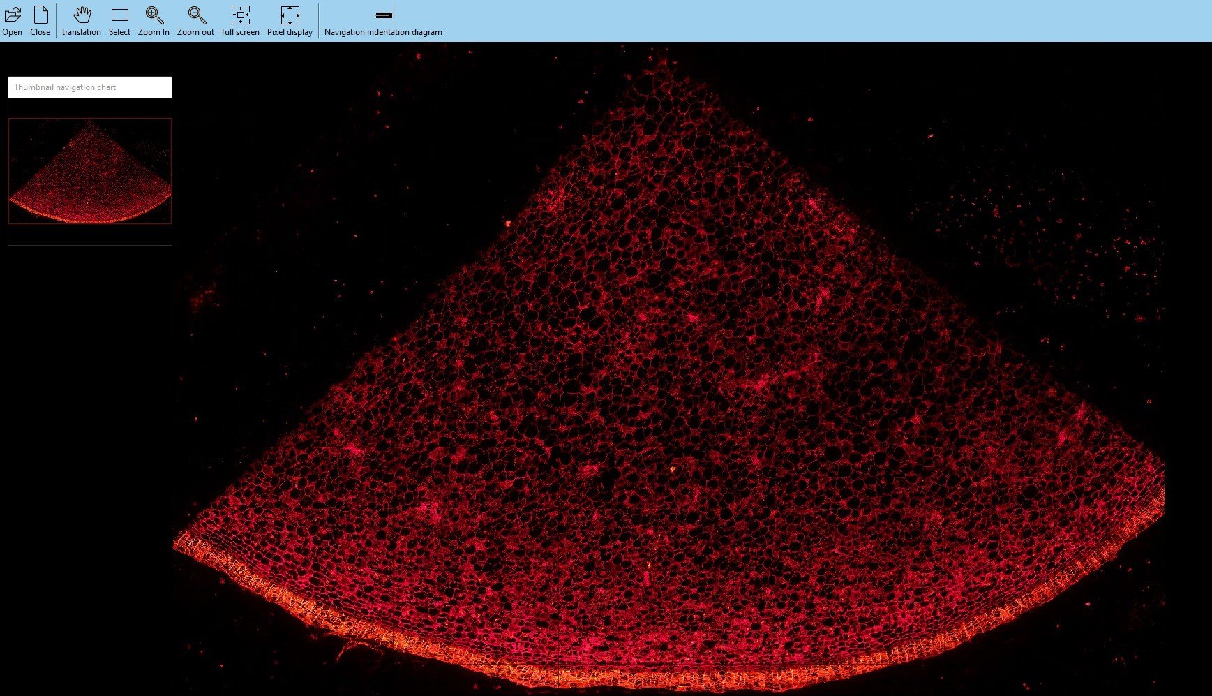
A 20x22mm area is scanned by 2.5x obj lens with a 5×8 tile and processed in less than 94 seconds (1min 34 sec). Below is the raw image with tiled boarder lines.
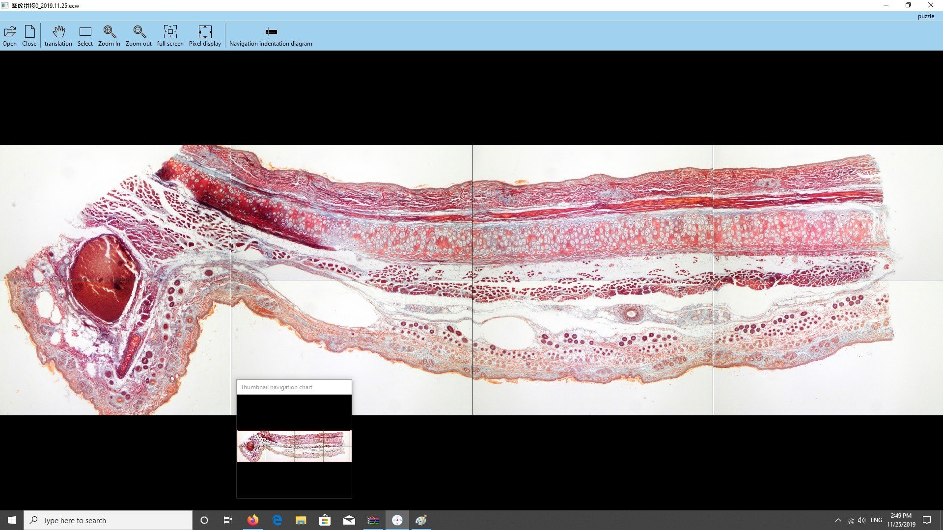
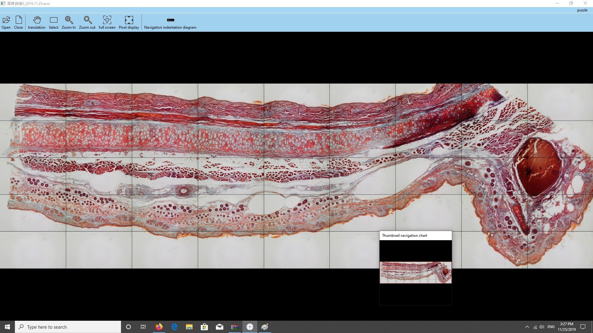
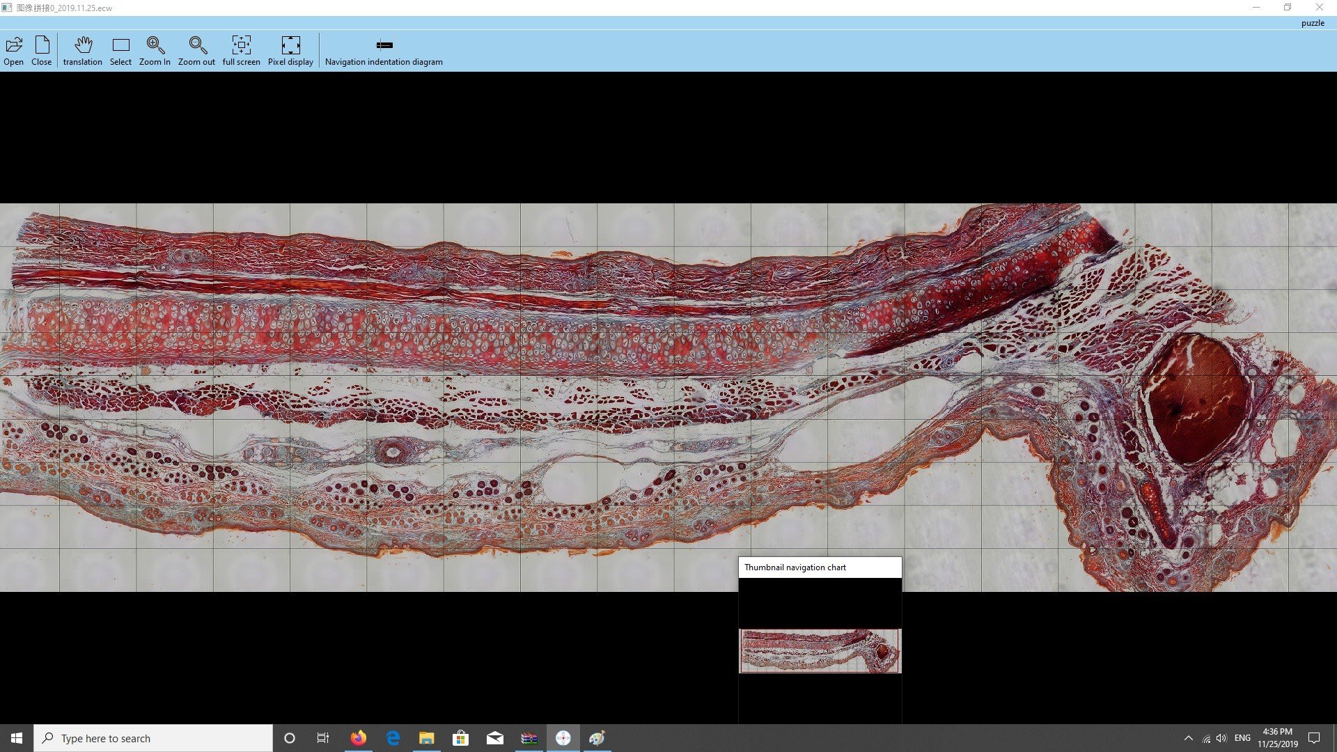
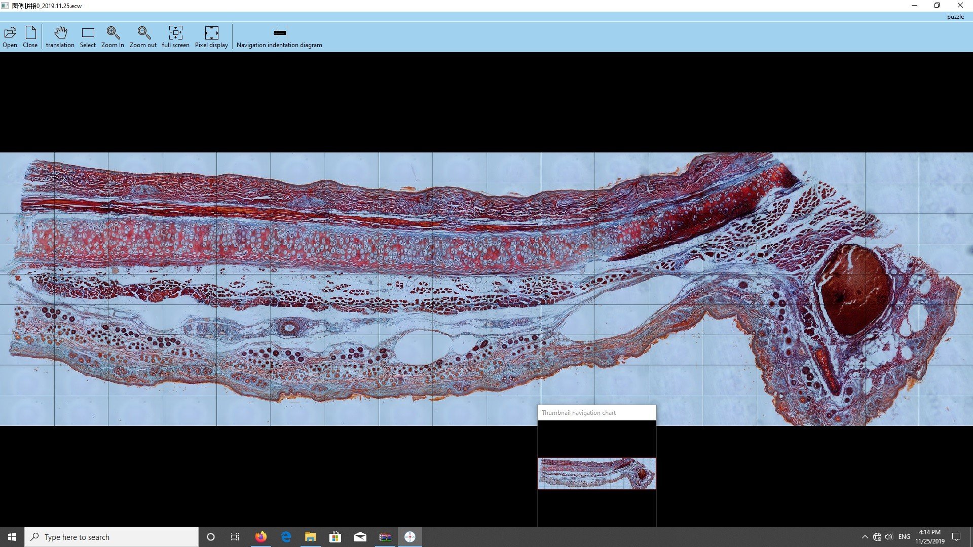
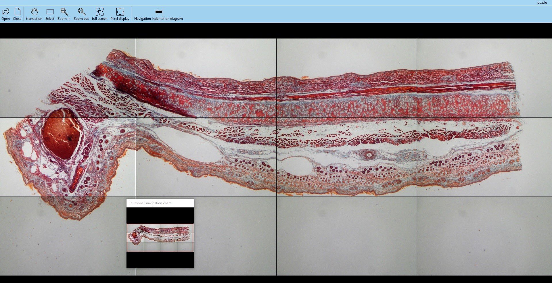
Example 1. Brightfield Imaging of a cartilage tissue slide
1.a. Using a 4x obj lens, and 5×3 tile:
1.b. Same Slide with 10x obj lens and 9×5 tiles
1.c. Cartilage Slide scanned with 20x obj lens, 18×9 tiles, manual exposure, autofocus every 5 spots
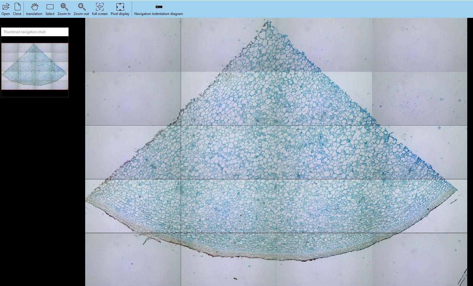
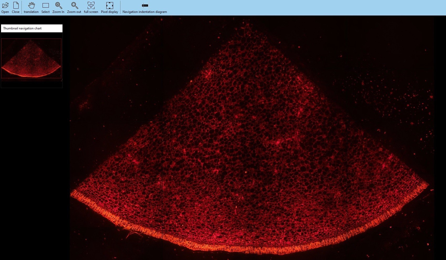
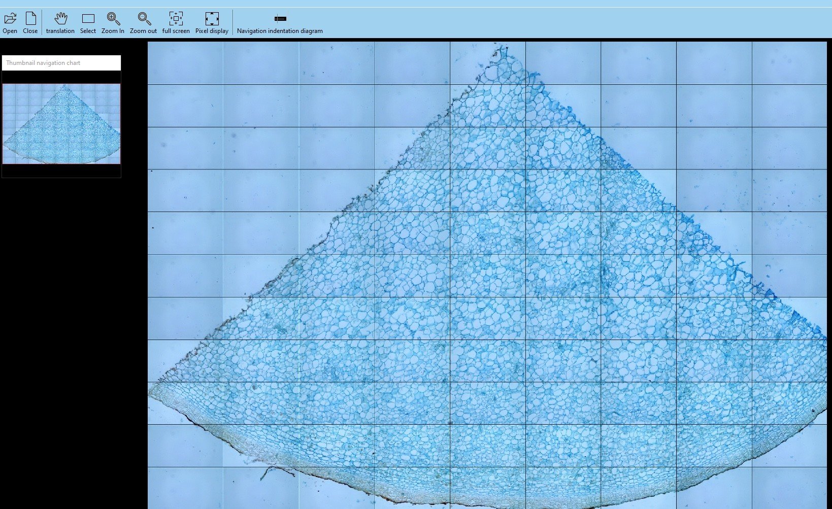
Example 2. Potato thin Section Slide
2.1. Brightfield imaging using 4x objective lens, with 4×5 tiles, autoexposure, non autofocus.
2.2. Fluorescence imaging using 4x objective lens, with 4×5 tiles, manual exposure 600ms, autofocus on every 3 spots with focus range of 10 steps of 40=400 steps (each step has 0.31um height).
2.3. Brightfield imaging using 10x objective lens, with 9×11 tiles, autoexposure, autofocus on every 3 spots with focus range of 16 steps of 8= total 128 steps (each step has 0.31um height).
2.4. Fluorescence imaging using 10x objective lens, with 9×11 tiles, autoexposure, autofocus on every 1 spots with focus range of 16 steps of 8= total 128 steps (each step has 0.31um height).
2.5. Fluorescence imaging using 20x objective lens, with 19×20 tiles, manual exposure, non autofocus.
Software
1. User friendly interface
The slide scanning software has a very easy to use and user friendly graphical user interface (GUI):
2. Easy and Simple Automation
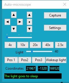
The software has a very simple interface. You can move the XY stage, Z focus, autofocus at three speeds of movement (low, medium and high) with a click of mouse. It automatically rotates the obj lens, shows the light intensity, coordinates of the current position (X,Y,Z) and saves 3 custom positions for scanning, start or stop.
3. Move and position your sample
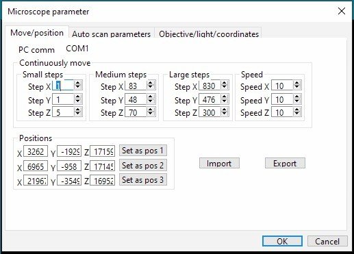
A sample can be moved and positioned at three different speeds of low, medium and high which can be set at custom values in all directions of horizontally (X), vertically (Y) and in height or depth of focus (Z).
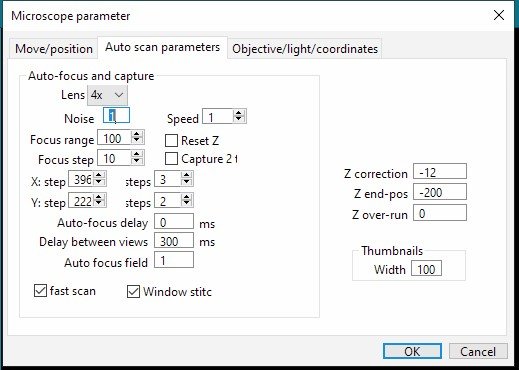
4. Automatic Scanning
Depending on which objective lens is selected, you can define, select and save the speed of movement, focusing range (number of focus, step size), step size (in X and Y direction), number of tiles (in X and Y), number of autofocusing, a pause time at each field of view.
You can select fast scan without autofocusing.
You can also determine to reset the focus when you rotate the nosepiece and move between objective lenses or shut down the system.
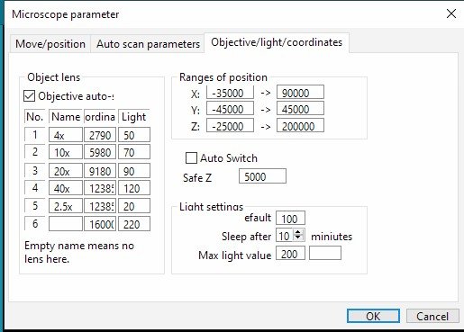
5. Custom Objective Lens name, Light Intensity and Coordinates
The software allows easily to rename the objective lenses, change their position, define a default light intensity for that specific objective lens. In below example, we often switch between 2.5x and 40x. The system remembers that is the same spot but it should apply a different light intensity.
For safety reasons, you do have access to change the limit of movements in all directions of X, Y and Z. You can also choose to release or not the focus height during the rotate of the nosepiece.
Please select the products based on your requirements below table:


Reviews
There are no reviews yet.