Live Cell Imaging
Showing 1–9 of 31 results
-
-
 Request a Quote Request a Quote
 -
$USD 1,999.00 Add to cart
-
$USD 3,995.00 Add to cart
-
-
-
 Request a Quote Request a Quote
 -
-
Showing 1–9 of 31 results
Live Cell Imaging
Live cell imaging is the study of the life of living cells / organisms using a time-lapse video microscopy in which the cells are kept at the closest conditions to a regular laboratory incubator. Live cell imaging is used by scientists and researchers to obtain a better understanding of dynamic events occur at cellular level under microscope.
What do you need to have to be able to get clear, stable and long-term images of your cells?
There are two approaches:
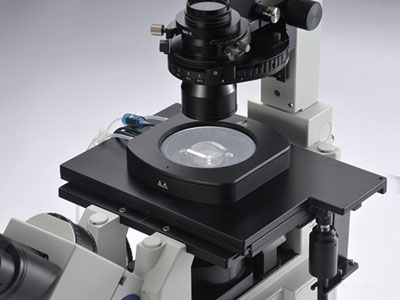
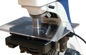
1. Use a classical microscope, camera and mount a miniature incubator / chamber on the microscope and also provide a gas, temperature and humidity;
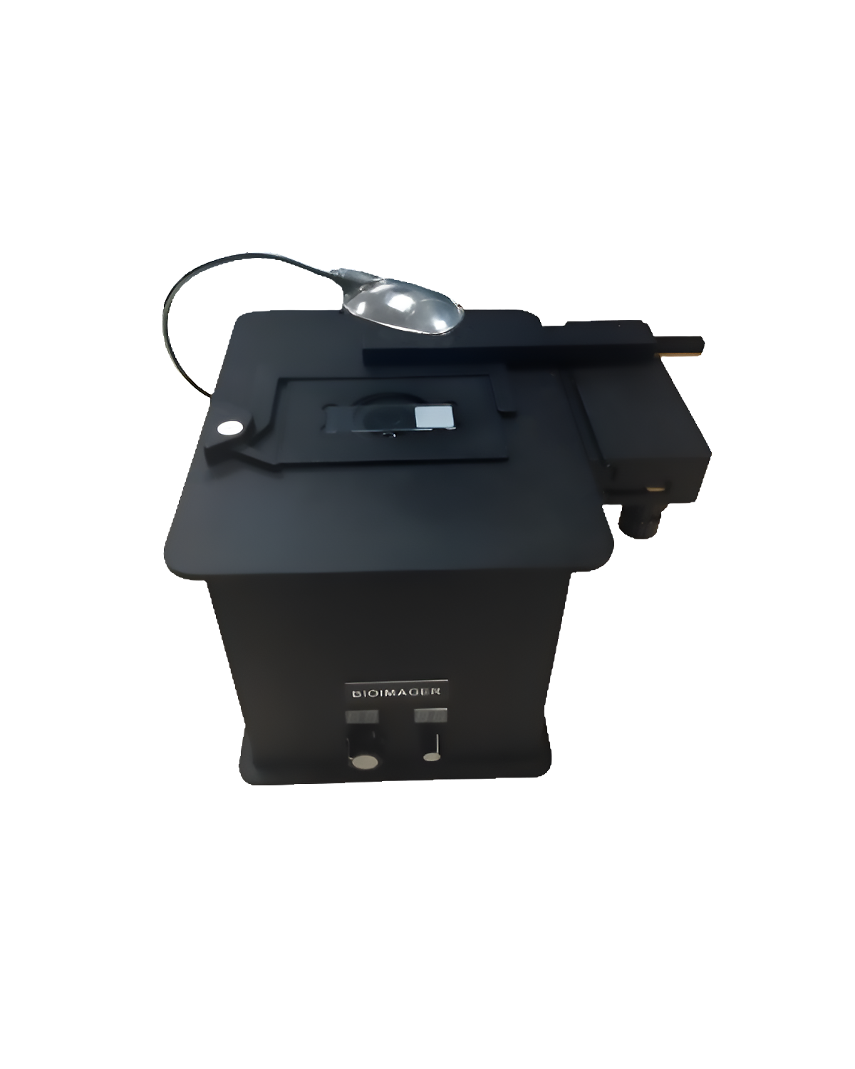
2. Use an Incubator Microscope which is small enough to keep inside a laboratory incubator, for instance IncuScope.
For most of the live cell imaging using the classical method, you will need at least these items beside a great microscope and camera:
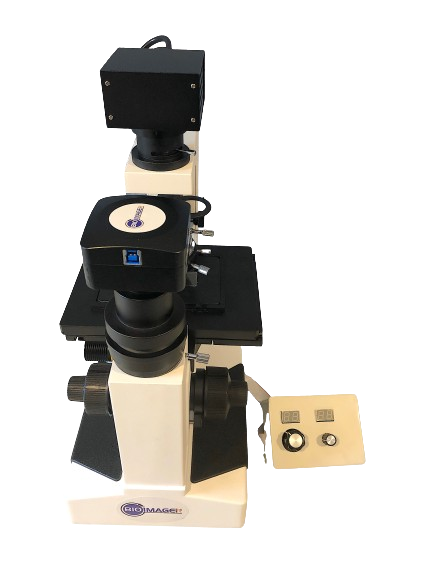
- A microscope
- A Camera
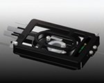
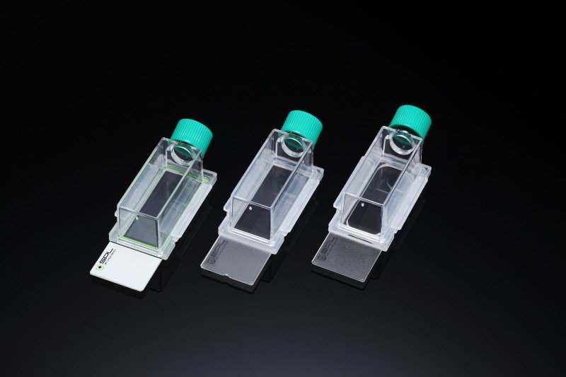
- A physical chamber, in which you can culture your cells. The chamber should allow easily installation and removal of the cell culture in a container such as well-plate, petri-dish, tissue culture flasks, micro-slide etc.
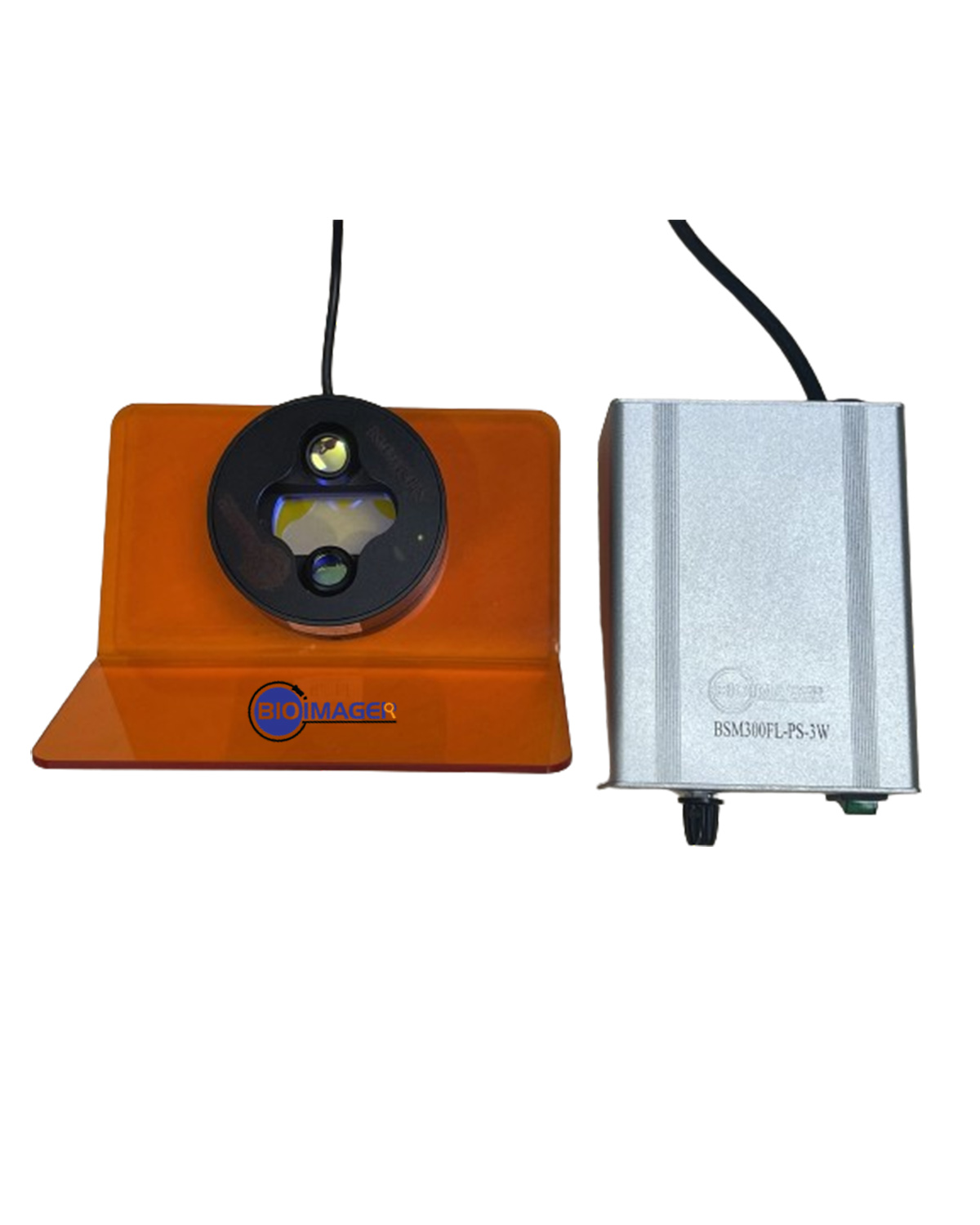
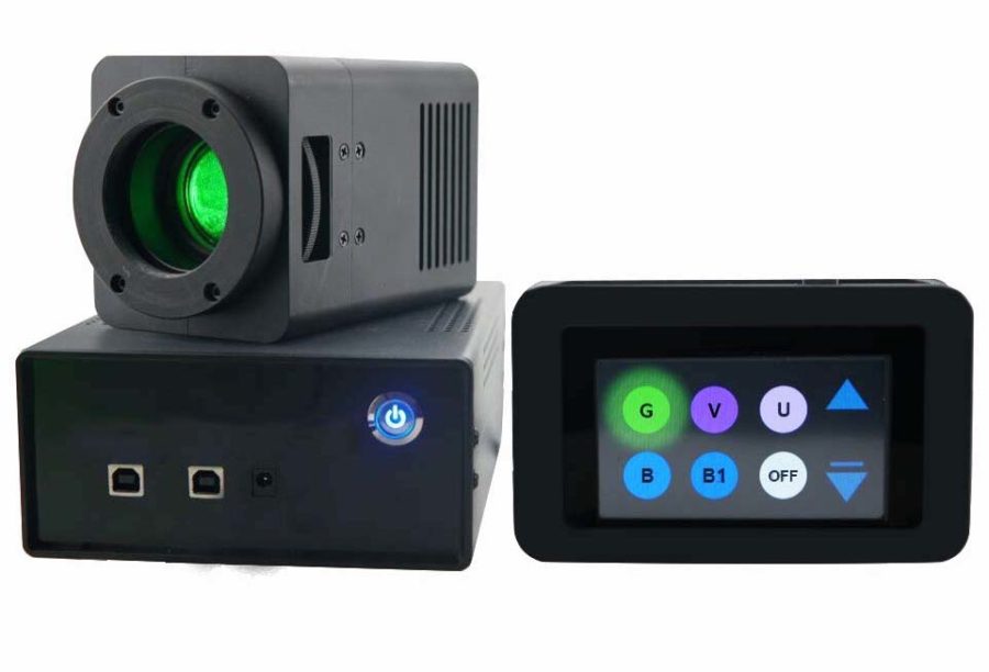
- A gas supplier, which can be a digital or analog gas mixer which provides the gas for your cells. The types include a mixed of CO2 and air, and mixed of CO2, O2, and N2.
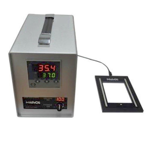
- A temperature controller, which allows to control temperature inside the chamber, outside the chamber and also surrounding the objective lenses to avoid drift focus (otherwise you will see blurry images due to ambient temperature fluctuation over time).
- A gas humidifier, this is simply a flow of premixed gas passes through water in a closed system.
- A glass-bottom dish or plate
- Lens Heater, to keep the lens not affected from the room temperature fluctuations.
- Shutter, to block the light if you do not need it during the waiting time and also avoid photobleaching and phototoxicity.
- A phenol-free medium
Beside these, generally speaking to obtain clear, stable, and long-term images of cells, you would need the following:
- A high-quality microscope: A good microscope is crucial for obtaining high-quality images. The microscope should have a high-resolution objective lens, a stable stage, and appropriate imaging software.
- Proper sample preparation: Proper sample preparation is essential for obtaining clear images of cells. This includes fixing the cells, staining them with appropriate dyes, and mounting them on a suitable medium.
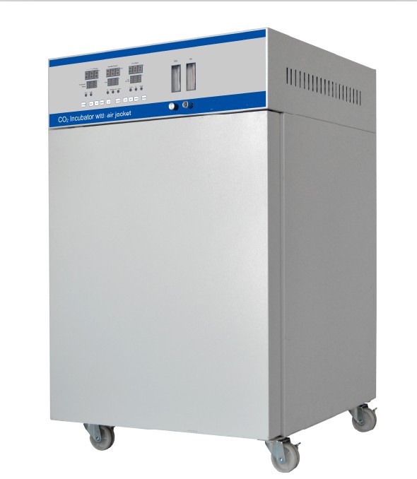
- A stable imaging environment: A stable imaging environment is important for obtaining stable images over a long period. This includes maintaining a constant temperature, humidity, and CO2 concentration.
- A suitable imaging technique: There are different imaging techniques available, such as brightfield, phase contrast, fluorescence, and confocal microscopy. The imaging technique used depends on the sample type and the desired imaging outcome.
- Quality imaging reagents: Using high-quality imaging reagents such as fluorescent dyes, antibodies, and probes can help improve the quality and specificity of the images.
- Careful handling of the sample: Careful handling of the sample is important to prevent damage to the cells and maintain their integrity during imaging.
- Experienced personnel: Experienced personnel who are familiar with the imaging technique and the microscope are essential for obtaining clear, stable, and long-term images of cells.
By taking into account all of the above factors, you can obtain high-quality images of your cells that are clear, stable, and can be used for long-term analysis.
In this category, you can find Live Cell Imaging Chambers, Miniature Incubators,Heating stage, Digital gas mixer, Temperature control, Objective lens heater, etc; Customized for all brands of fluorescent & confocal microscopes.
Still have a question, use this Live Cell Imaging Selection Questionnaire.