Darkfield
Showing 10–15 of 15 results
Showing 10–15 of 15 results
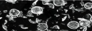
A darkfield biological microscope is a type of optical microscope that uses specialized illumination techniques to enhance the contrast of transparent, unstained specimens. In a darkfield microscope, the sample is illuminated with a hollow cone of light that does not enter the objective lens directly. Instead, the light is scattered by the sample, causing it to appear bright against a dark background.
The dark background in a darkfield microscope is achieved by blocking the central portion of the light beam, allowing only the peripheral scattered light to reach the objective lens. This technique effectively eliminates the light that would normally be scattered directly by the sample, producing a bright image of the sample against a dark background.
Darkfield microscopy is commonly used in biological research and medical diagnosis to observe and study bacteria, parasites, and other small, transparent organisms that are difficult to see with other types of microscopes. Darkfield microscopy is also useful for studying living samples, as it does not require staining or fixation of the sample, which can alter its natural characteristics.
Overall, darkfield microscopy is a valuable tool for studying transparent and difficult-to-see samples, and it is commonly used in a variety of fields, including microbiology, cell biology, and medical research.
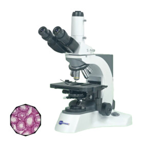
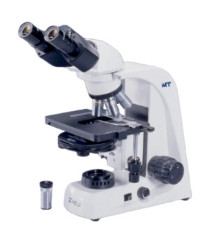
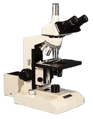
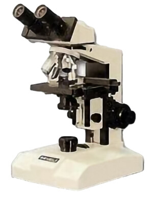
Biological microscopes with darkfield condenser and dark-field objective lenses are great for live or dead blood cell analysis with great drak-ground or dark background images. It is a low-price alternative to phase-contrast imaging.

