Introduction
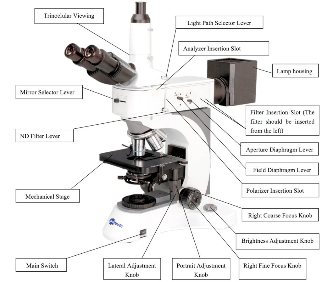
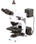
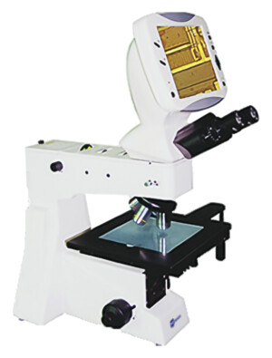
BMU500DIC RF/TRF metallurgical microscopes are high level professional microscopes which are specially designed for metallurgical analysis. With excellent optical system, ingenious stand and convenient operation, they will be your best choice. DIC observation attachment is optional for these microscopes to meet some special requirements.
Application
BMU500RF/TRF series are widely used in institutes and laboratories to observe and identify the structure of various metal and alloy, they also can be widely used in electronics, chemical and instrumentation industry, observe the opaque material and transparent material, such as metal, ceramics, integrated circuits, electronic chips, printed circuit boards, LCD panels, film, powder, toner, wire, fibers, plated coatings, and other non-metallic materials and so on.
Features
1. Laboratory metallurgical microscope, including bright field, dark field, polarization and DIC observation system.
 2. Powerful transmitted and reflected system with Kohler illumination.
3. Ideal instrument for metallurgical analysis, industry inspection and science research.

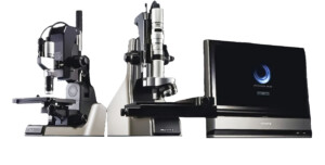
BMU500DIC Metallurgical Microscope with BF/DF and optional Polarized and DIC Nomarski Imaging
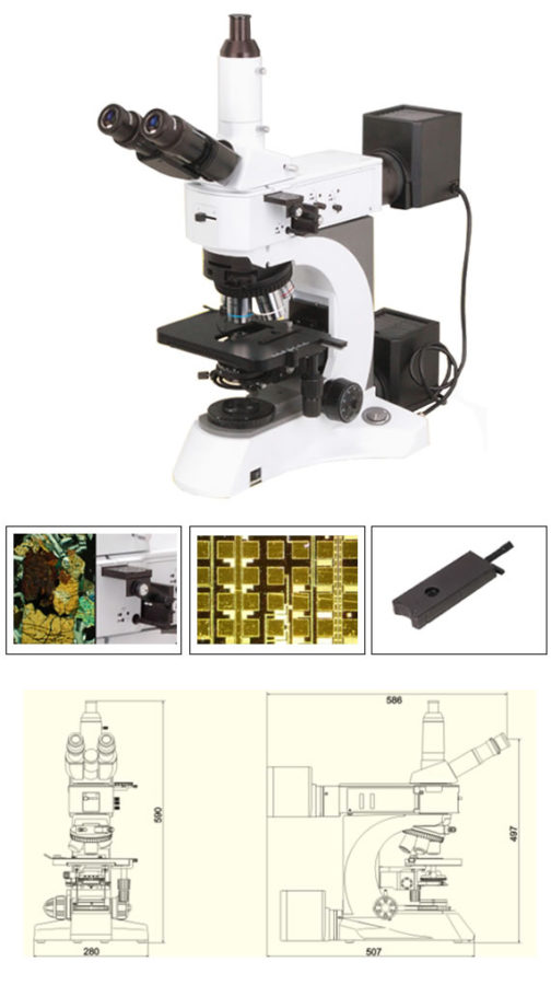
BMU500DIC Laboratory DIC BF/DF Metallurgical Microscope, is an ideal instrument for industry inspection and science research laboratory. –Infinity Optical System, BF/DF, Transmit & Reflect Illumination, DIC Nomarski, Polarizing System –Full Range BF/DF Objectives Included –Polarization System Optional –DIC System Optional –Powerful Transmitted And Reflected Illumination System with Kohler Illumination.
| Model |
BMU500DIC
RF |
BMU500DIC
TRF |
| Optical System |
Infinite optical system |
● |
● |
| Viewing Head |
Siedentopf trinocular viewing head, inclined at 30°, interpupillarty distance 48mm-75mm |
● |
● |
| Eyepiece |
Extra wide field eyepiece EW10×/22, eyepiece tube Φ30mm |
● |
● |
| Infinite Plan Achromatic Objective |
5×/ 0.12/∞/ – (BF/DF) WD 12mm |
● |
● |
| 10×/ 0.25/∞/ – (BF/DF) WD 10.0mm |
● |
● |
| 20×/ 0.4/∞/ 0 (BF/DF) WD 4.3mm |
● |
● |
| 40×/ 0.65/∞/ 0.17, WD 0.54mm |
|
● |
| 100×/ 1.25/∞/ 0.17, WD 0.13mm |
|
● |
| 40×/ 0.6/∞/ 0 (BF/DF) WD 2.9mm |
○ |
○ |
| 50×/ 0.75/∞/ 0 (BF/DF) WD 0.32mm |
● |
● |
| 100×/ 0.8/∞/ 0 (BF/DF) WD 2mm |
● |
● |
| DIC |
20×, 100× |
○ |
○ |
| Maximum Sample Height |
30mm |
|
● |
| 50mm |
● |
|
| Reflected Light |
24V/100W Halogen light, Brightness adjustable |
● |
● |
| Kohler illumination and aspherical condenser |
● |
● |
| Polarizer and analyzer |
○ |
○ |
| An integrate device for polarizer and analyzer |
○ |
○ |
| Blue, Green, Yellow and Frosted filter |
● |
● |
| Transmitted Light |
Swing-out condenser NA0.9/ 0.25 |
|
● |
| 24V/100W Halogen light and aspherical condenser |
|
● |
| Blue filter |
|
● |
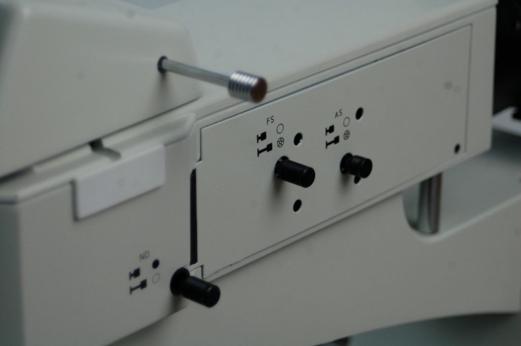
| Filter |
ND25, ND6 |
○ |
○ |
| Focusing |
Coaxial Coarse and Fine Adjustment, Fine Division 0.001mm, Coarse Stroke 37.7mm per Rotation, Fine Stroke 0.1mm per Rotation, Moving Range 24mm |
● |
● |
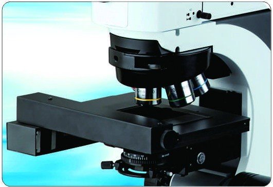
| Nosepiece |
Backward quintuple nosepiece |
● |
● |
| Stage |
Double layer mechanical stage(without hole) 186×138mm/ 74mm×50mm |
● |
|
| Double layer mechanical stage 186×138mm/ 74mm×50mm |
|
● |
| Glass specimen preparation plate |
○ |
○ |
| Specimen preparation plate |
|
● |
| Slide glass |
|
● |
| Accessories |
Specimen Presser |
○ |
○ |
| Photo Attachment |
○ |
○ |
| Video Attachment, C Mount 1×, 0.5× |
○ |
○ |
Note: ●Standard Outfit, ○Optional
Video 1: Main Body and Performance Overview:
Video 2: Imaging Capability: Brightfield imaging with transmitted or Reflected Light
Video 3. Using 16MP to show transmitted brightfield sample images of BMU500
This video demonstrates the microscopy camera BRC-1600 which is a 16MP 1/2.33″ USB 3.0 colorful camera. The camera is connected to one of our upright industrial /metallurgical microscope BMU500DIC with 0.37x c-mount adapter, Windows 10, and sample images of Tilia (Basswood) are shown at different objective lenses.
Video 4. Fluorescence Imaging
This video demonstrates Bioimager BUM360FLED-A LED Fluorescence attachment to use with a standard upright microscope such as BMU500DIC. It comes with 5W LED of Violet (V), Ultraviolet (UV), Blue (B) and Green (G) LEDs, and its correspondent excitation and emission filters. The video demonstrates the optics and mechanics as well as sample images at different magnifications.
Custom Microscopy package of this model was provided to several customers. These are some examples:
- Thorlabs Quantum Electronics, MD, USA
- Montana State University, Bozeman, MT, USA
- POSTECH University, S. Korea
- A private corporate at Cabrière d’Avignon, France
- International Sci. & Technol. Center (ISTC) Astana, Kazakhstan & Yerevan, Armenia
- A private firm at Brownstown, MI, USA
Only logged in customers who have purchased this product may leave a review.


Reviews
There are no reviews yet.