Automated Live Cell Imaging – Enables real-time monitoring with bright-field and fluorescence microscopy.
Multi-Position Imaging – Fully motorized system captures images across multiple points, supporting up to 96 wells.
Z-Stack Imaging – Captures multiple focal planes for enhanced depth and clarity.
Compact & Incubator-Compatible – Designed to fit inside standard CO₂ incubators without disrupting cell culture conditions.
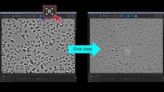
Autofocusing System – Ensures fast, precise, and reproducible focus adjustments.
Image Stitching Function – Seamlessly merges multiple images for large-scale cell analysis.
High Compatibility – Supports various cell culture vessels including flasks, dishes, well plates, and slides.
User-Friendly Software – Provides confluency measurement, growth curve analysis, and digital ruler tools.
Ideal for Cell-Based Research – Suitable for cell proliferation studies, wound healing assays, apoptosis analysis, drug discovery, and high-throughput screening.
Live cell imaging is an advanced microscopy technique used to observe living cells over time using time-lapse microscopy. This method enables researchers to analyze cellular processes in real time without disrupting their natural state.
Key Benefits of Live Cell Imaging:
Essential for Life Science Research – A fundamental tool in cell biology, drug discovery, and medical research.
Understanding Cellular Dynamics – Helps study biological processes such as cell division, migration, and interactions.
– Reduces handling errors and preserves cell viability for accurate analysis.
Delivers Reliable Data – Provides continuous observations, offering deeper insights compared to endpoint analysis.
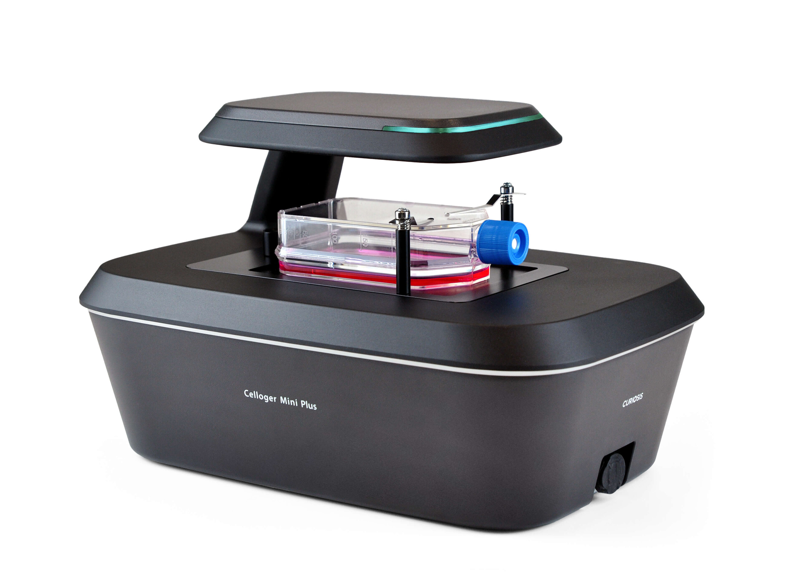
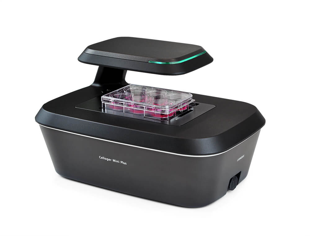
Celloger Mini Plus – Advanced Automated Live Cell Imaging System introduction:
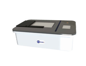
Celloger® Mini Plus is an advanced automated live cell imaging system designed to enhance real-time cell monitoring, analysis, and research applications. Equipped with bright-field and fluorescence microscopy, it enables high-resolution imaging with autofocusing, multi-position imaging, and Z-stack functionality.
With its compact and portable design, Celloger® Mini Plus seamlessly integrates into standard CO₂ incubators, allowing continuous observation without disturbing the optimal cell culture environment. The system supports various cell culture vessel types, making it ideal for cell proliferation studies, wound healing assays, apoptosis analysis, drug discovery, and high-throughput screening.
By combining cutting-edge imaging technology with user-friendly software tools for confluency measurement, growth curve analysis, and image stitching, Celloger® Mini Plus ensures precise, reliable, and reproducible results for biomedical research, pharmaceutical development, and regenerative medicine.
Celloger Mini Plus –Advanced Live Cell Imaging – Key Features & Benefits
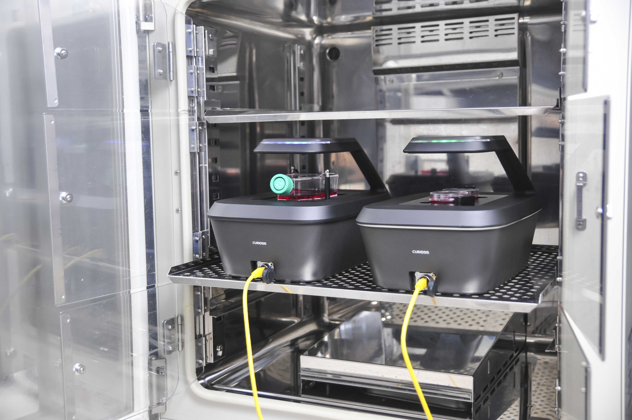
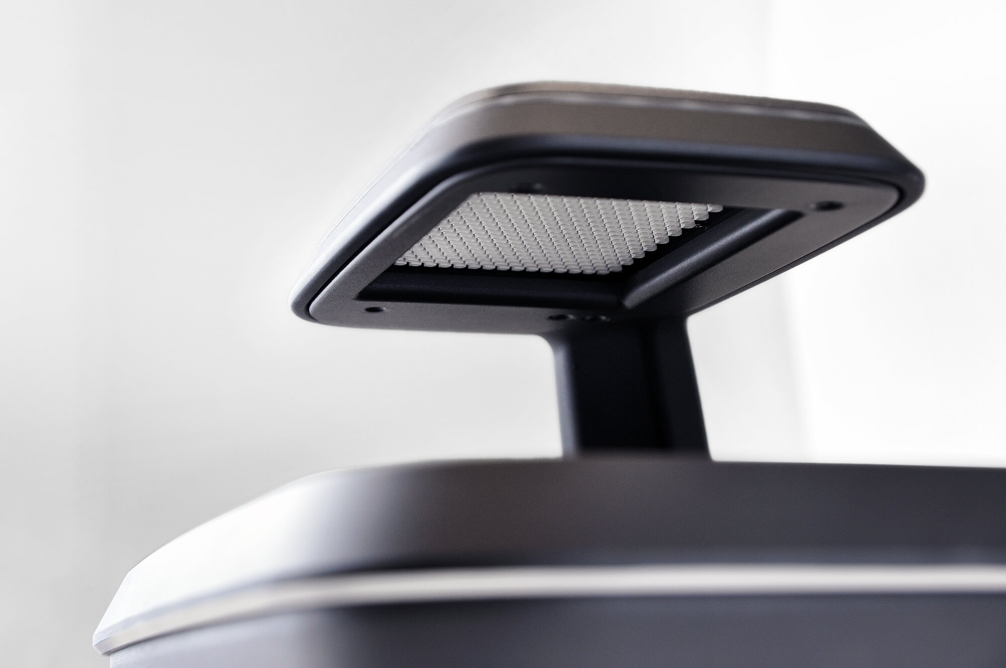
1. Real-Time Cell Monitoring with Optimized Environmental Stability
Monitoring live cells in hot and humid incubator environments presents a significant challenge, as fluctuations can affect cell viability and experimental accuracy. The Celloger® Mini Plus is designed to operate seamlessly inside the incubator, allowing long-term, real-time cell observation without disrupting the optimal conditions required for cell culture growth. By eliminating the need to remove samples, it minimizes contamination risks and ensures consistent, high-quality imaging for reliable research outcomes.
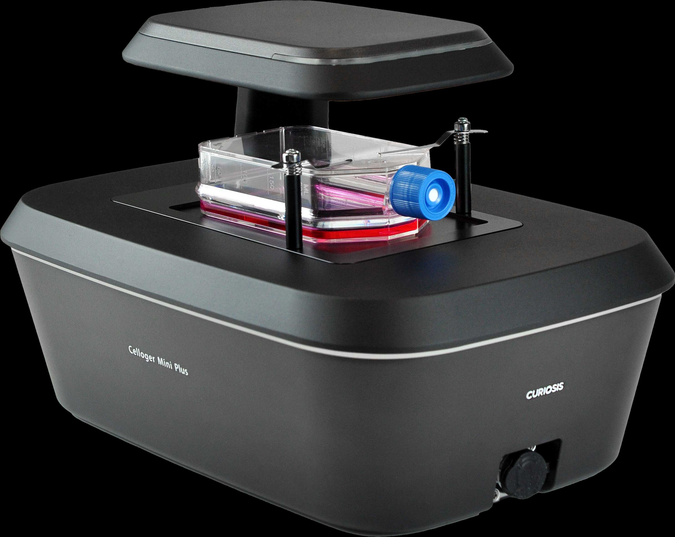
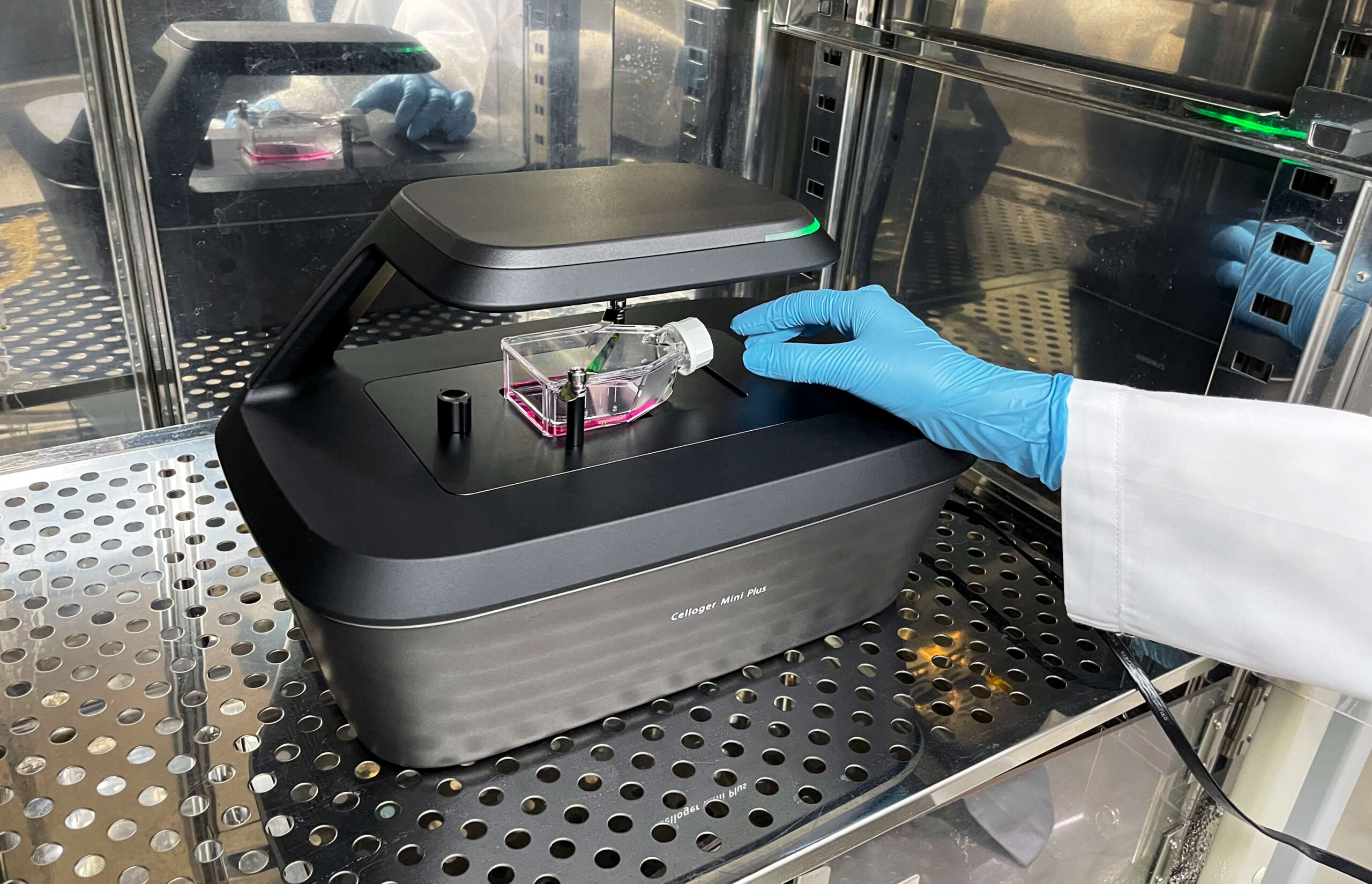
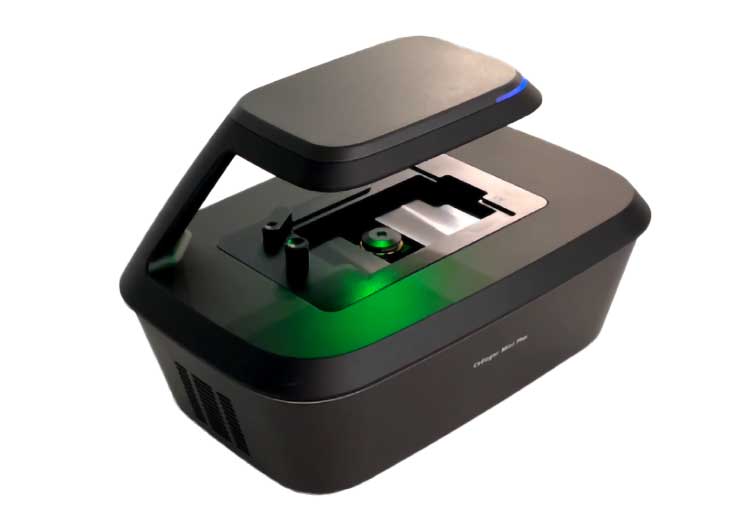
2. Compact & Portable Design for Flexible Lab Integration
The Celloger® Mini Plus is engineered with a space-efficient design (226H × 358L × 215W mm), allowing multiple units to fit seamlessly inside a standard CO₂ incubator. Weighing approximately 5.6 kg, this lightweight system is easy to transport in and out of the incubator as needed, offering maximum flexibility for dynamic research environments. Its compact footprint ensures it occupies minimal lab space while maintaining high-performance live cell imaging capabilities.
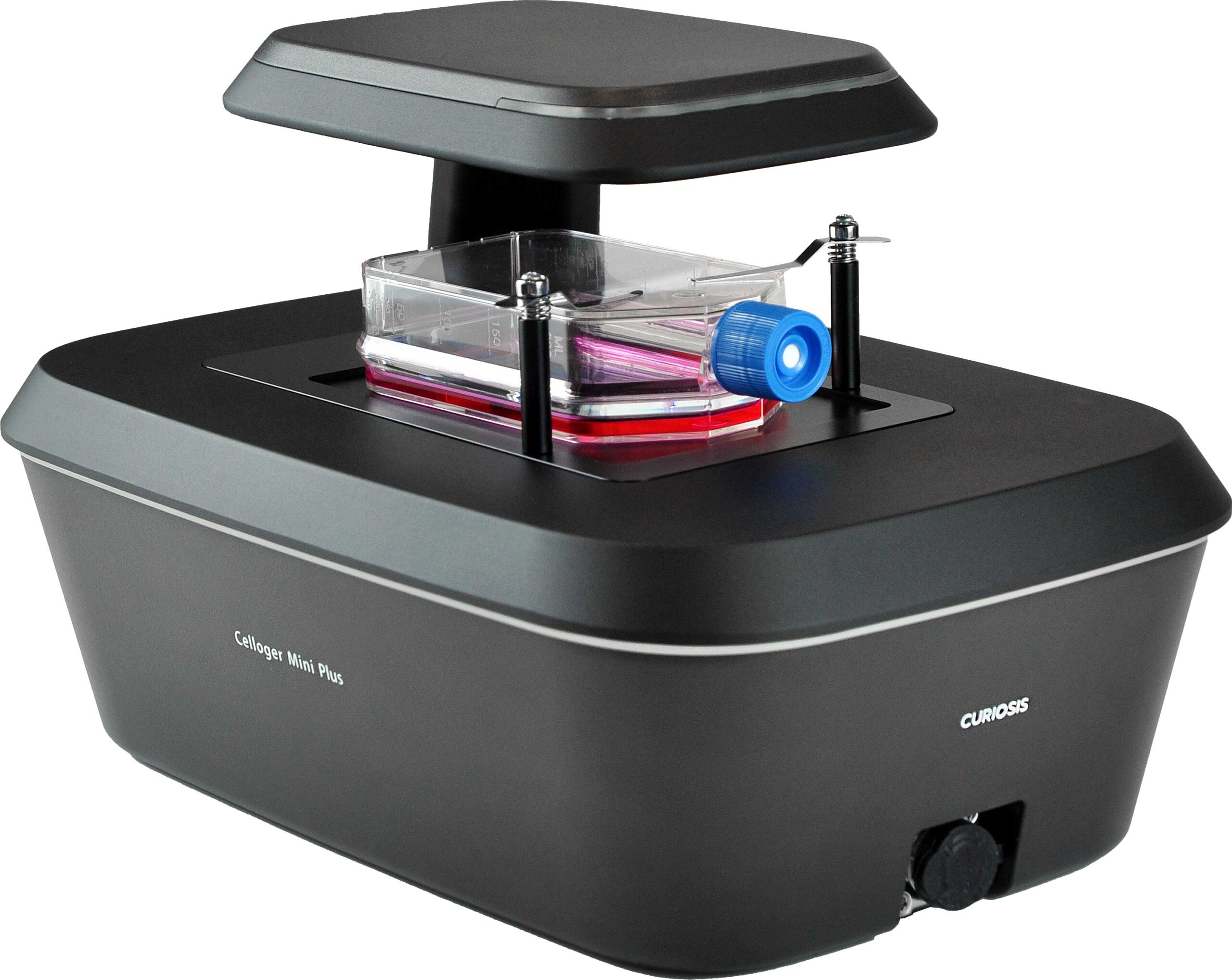
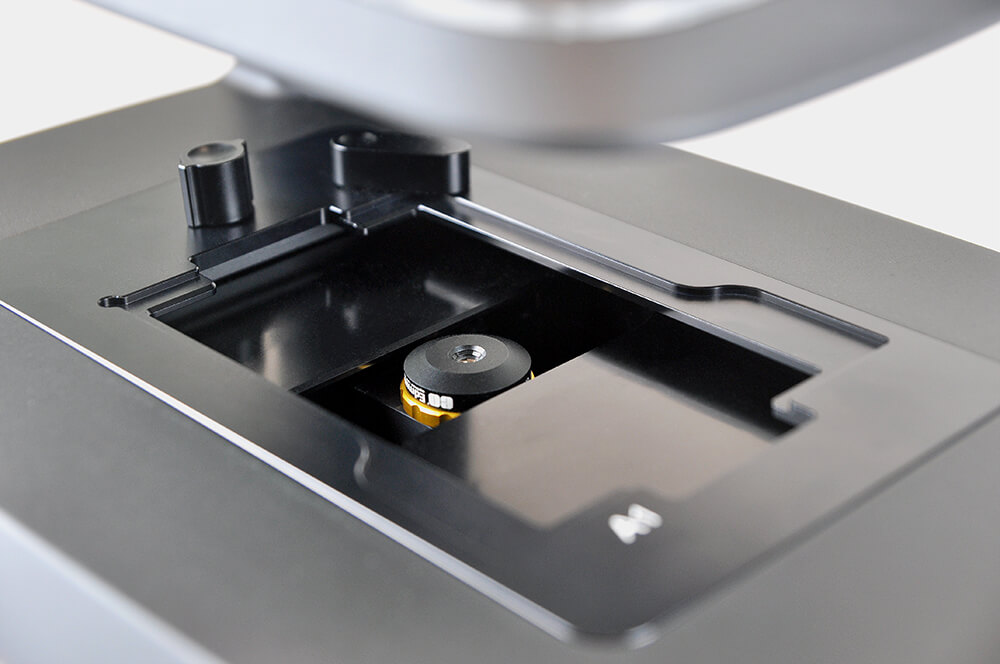
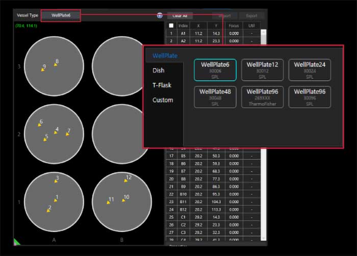
3. High Compatibility with Cell Culture Vessels
Designed to support a wide range of cell culture vessel types, including flasks, dishes, well plates, and slides, making it ideal for diverse cell-based research applications.
Celloger® Mini Plus – Advanced Automated Live Cell Imaging System
Celloger® Mini Plus is a high-performance automated live cell imaging system designed for precise and reliable research. Featuring advanced fluorescence and bright-field microscopy, autofocusing, and real-time multi-position imaging, it ensures high-resolution image acquisition with superior accuracy.
This all-in-one system supports a wide range of cell-based research applications, providing essential tools for high-quality imaging and reproducible results.
Enhance your cell research with Celloger® Mini Plus – the ultimate solution for automated live cell imaging.
Celloger Mini Plus – Advanced Automated Live Cell Imaging System Specification
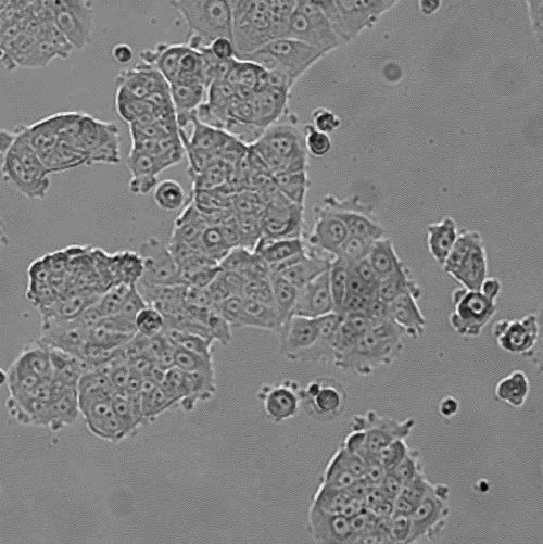
Applications of Live Cell Imaging – Captured with Celloger® Mini Plus:
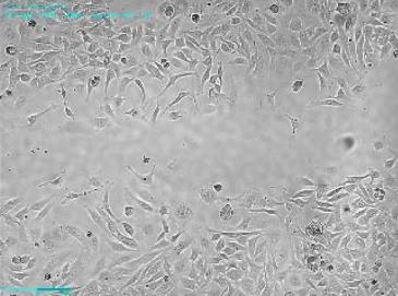
Cell monitoring:
Live cell monitoring minimizes labor time and contamination risks by enabling real-time observation of cell growth without disrupting culture conditions.
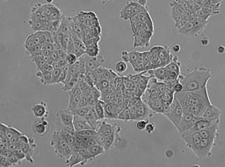
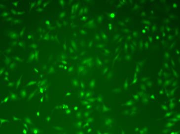
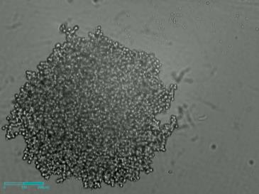
Cell proliferation:
Cell proliferation refers to the increase in cell numbers due to division. It can be visualized, quantified, and analyzed for research applications.
Wound healing assay:
This assay is widely used to study cell migration, cell polarization, and tissue repair across various cell types in research.
Coculture monitoring:
Coculture monitoring examines cell-to-cell interactions between two or more cell types using live imaging technology to understand cellular behavior.
Cytotoxicity:
Cytotoxicity assesses the toxicity of substances on cells, identifying how chemical or environmental factors suppress cell growth and affect cell health.
Apoptosis:
Apoptosis, or programmed cell death, involves key processes like membrane blebbing, cell shrinkage, and nuclear fragmentation, crucial for disease research.
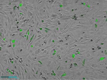
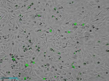
Reactive oxygen species(ROS) detection:
Excessive oxidative stress from ROS can lead to cell damage and disease progression, making its detection critical in cancer and neurodegenerative research.
Spheroid screening:
3D cell culture models help overcome the limitations of 2D cultures, better simulating real tissue environments for advanced research.
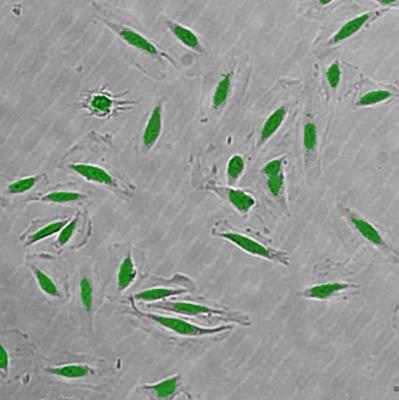
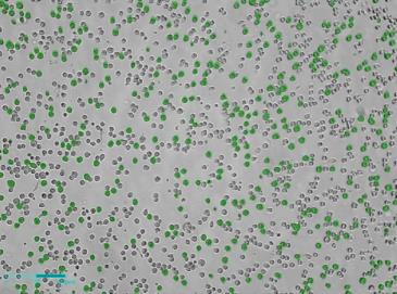
Transfection:
Transfection efficiency is measured to optimize gene delivery into cultured cells, ensuring high viability and successful gene expression.
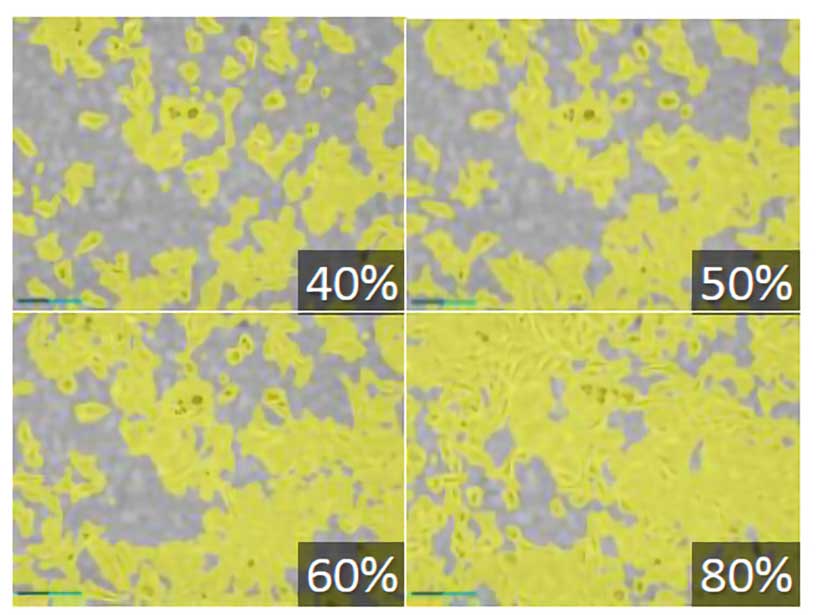
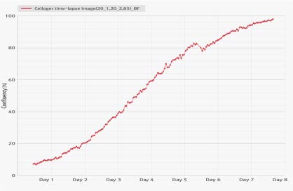
Cell culture & growth:
By analyzing the cell occupied area ratio (confluency), researchers can track cell growth trends and create graphs to quantify cell expansion over time.
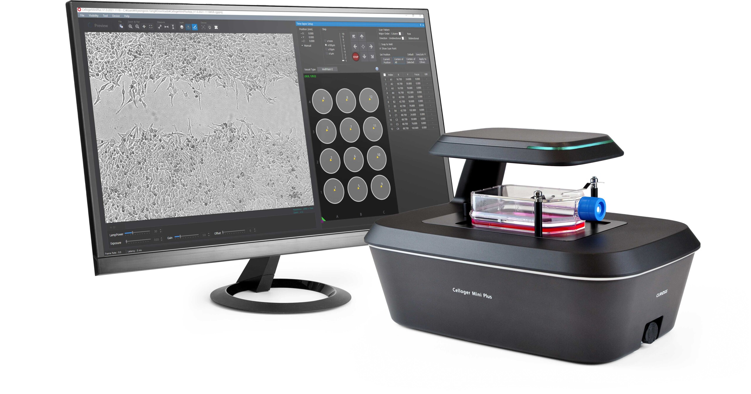
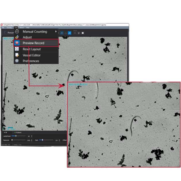
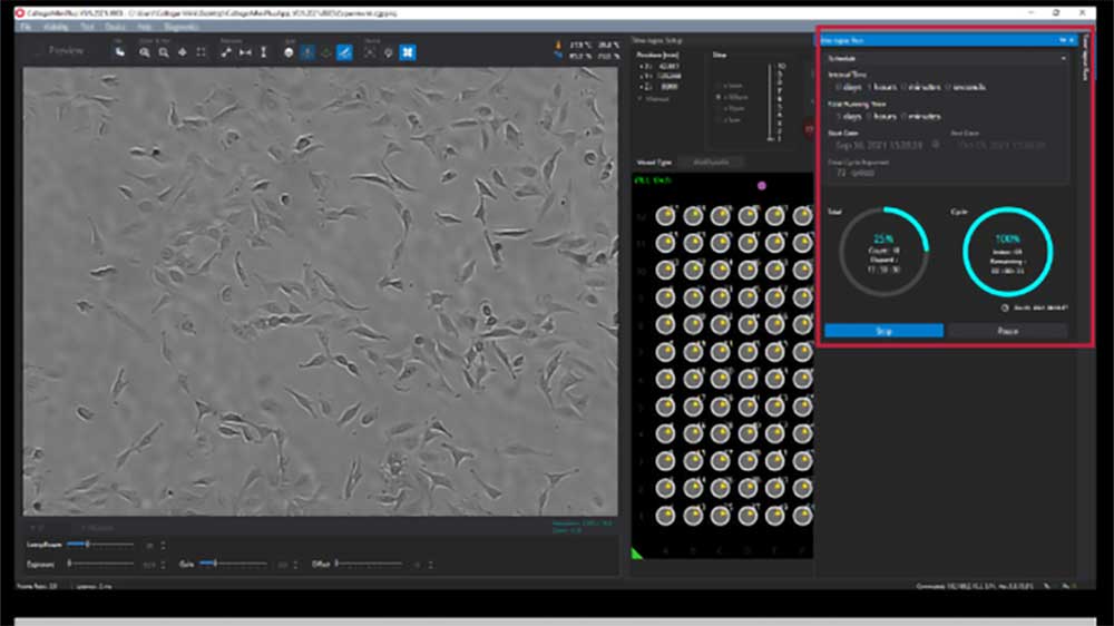
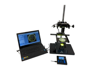
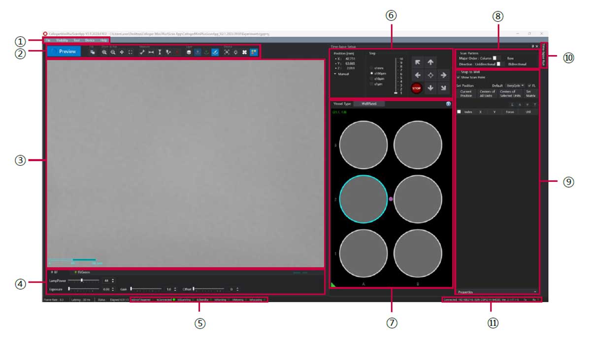
Software Overview
The Celloger® Mini Plus Scan App is an intuitive software designed to control the Celloger® Mini Plus device, enabling seamless real-time cell monitoring and time-lapse imaging. This powerful application enhances live cell research by providing automated imaging, precise data acquisition, and efficient workflow management for researchers.
①Menu bar
②Toolbox
③Display (Preview)
④Light source control
⑤Device status
⑥Jog control
⑦Vessel & Plate panel
⑧Scan pattern
⑨Position control
⑩Time-lapse control
⑪Connection status
Multi-Position Imaging for High-Throughput Studies
The Celloger® Mini Plus features a fully motorized camera system that enables precise multi-point imaging across an extended 117mm × 77mm travel range (X and Y axes). This advanced automation allows researchers to capture multiple points within the designated area at pre-set intervals, cycles, and total time, ensuring consistent, time-lapse imaging of live cells.
With the ability to image up to 96 wells, this system is an ideal solution for high-throughput screening, drug discovery, stem cell research, and cell-based assays. By automating multi-point image acquisition, it significantly enhances efficiency, reproducibility, and data accuracy in long-term live cell experiments.
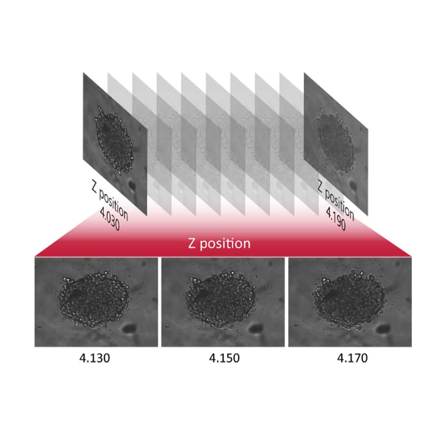
Z-Stack Imaging for Enhanced Focus
Capture multiple focal planes using advanced Z-stack imaging technology, providing deeper insights into cell morphology and structure.
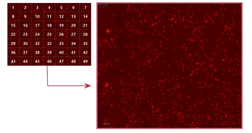
Image Stitching for Large-Scale Analysis
The automated image stitching function seamlessly merges multiple images, enabling researchers to analyze larger cell sections with high precision.
Intuitive & User-Friendly Software
Built-in cell analysis tools simplify research workflows with features like:
Confluency measurement for accurate cell growth tracking
Growth curve visualization for real-time progress analysis
Digital ruler & annotations for precise data documentation
Autofocusing for High-Resolution Imaging
Achieve faster and more reliable focusing with an intelligent autofocusing system, ensuring high-quality, reproducible live cell imaging results.
The Celloger® series is designed for use in a wide range of scientific research disciplines, playing a crucial role in the early stages of experimentation. Its advanced live cell imaging capabilities support researchers in academic institutions and industrial settings alike, with a balanced 50:50 distribution between both sectors.


Reviews
There are no reviews yet.