Phase Contrast MicroscopyPhase Contrast MicroscopyPhase Contrast Microscopy
What is Phase Contrast Microscopy?
Phase Contrast is a contrast enhancing optical microscopy technique in which an unstained transparent sample is seen with high brightness by shifting phase difference of light.
The idea of Phase Contrast Imaging was brought by Frits Zernike in 1930’s, a Dutch physicist, who got the Nobel prize in 1953 for this invention.
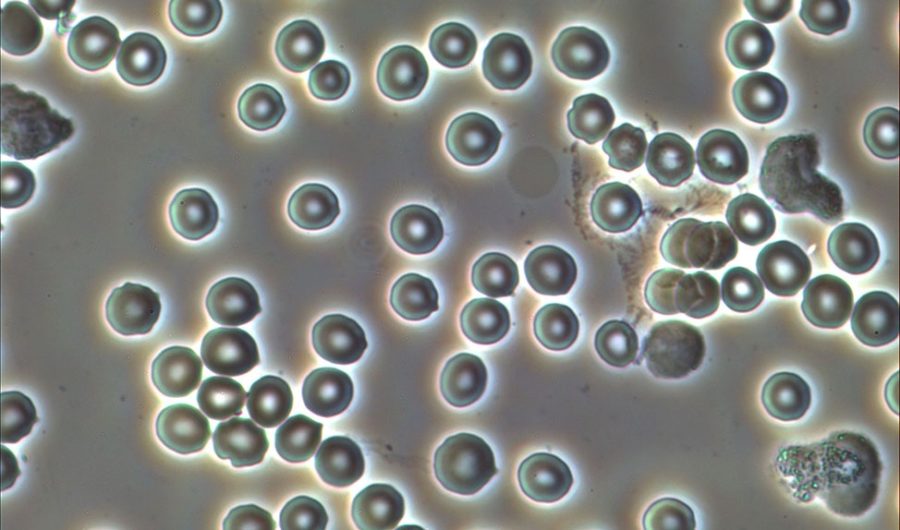
Phase contrast is mainly used by biologists and has some advantages over brighfield imaging. Using phase contrast microscopy, many cellular structures that cannot be seen by a brightfield imaging become visible, without staining.
Phase Contrats imaging is a great technique for live blood cell analysis
Phase Contrast Microscopes
Phase Contrast microscopes are mainly offered in biological / clinical microscopes and for transparent samples only. Opaque or non-transparent samples need DIC Nomasrky Imaging technique. Thus, biological microscope either upright or inverted comes with extra accessory for Phase Contrast imaging. This needs a condenser, phase contrast objective lens and annulus plate.
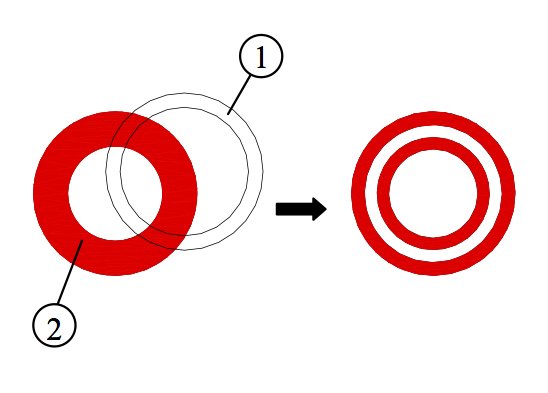
Key Optical Elements for Phase Contrast Microscopy
1. Phase contrast obj lens
Every phase contrast objective lens has a phase ring inside and is normally labeled with Ph sign which can be Ph1, Ph2, Ph3 or so. If you look at a phase contrast lens from the thread side, you will easily see such a ring. It is important to know that the phase contrast lens can be used for brightfield imaging but should not be used for fluorescence imaging as the fluorescence light damages the ring inside the lens and even may cause it melt.
It is important to know that the phase contrast objective lenses can be used for brightfield imaging but they should not be used for fluorescence imaging as the fluorescence light damages the ring inside the lens and even may cause the lens melt. This is a very common mistake that we hear from our clients.
2. Condenser
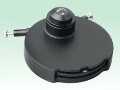
Upright Microscope
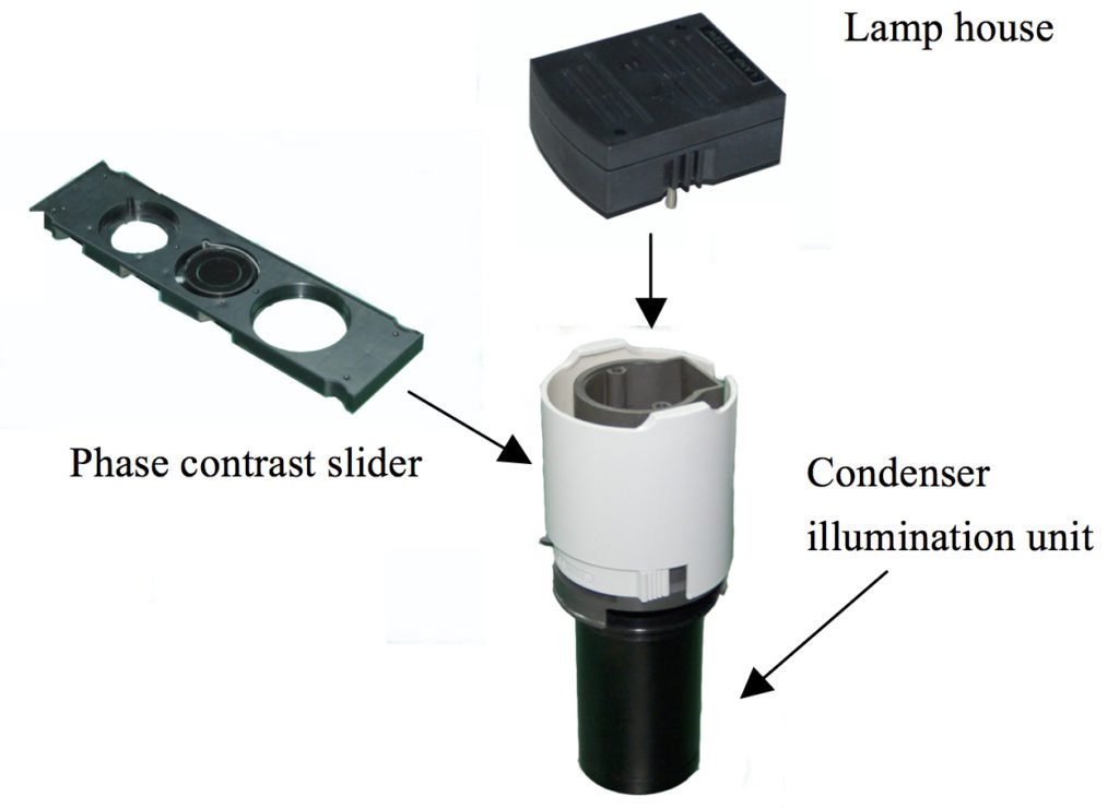
Inverted Microscope
3. Phase Annulus

How to align a phase contrast microscope?
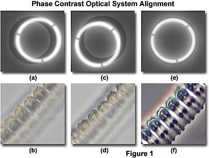
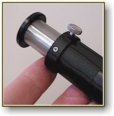
1. Basic Method: Use a phase contrast alignment tool
2. Intermediate Method: Use a piece of paper
3. Advanced Method: Work directly on sample
Check this video tutorial that reviews these methods:

