
Brightfield Image:
Brightfield (BF) is the simplest imaging technique of all the optical microscopy imaging mode. As the name states you can expect an image with bright background. A simple example in our daily life is like seeing birds or airplane in the sky. In BF imaging, a sample is normally illuminated via transmitted light coming from a white light and contrast in the sample is caused by the absorbance of some of transmitted light in dense areas of the sample.
Polarizing Image:
A type of optical microscopy techniques involving polarizing light. Simple techniques include illumination of the sample with polarized light. Directly transmitted light can, optionally, be blocked with a polarizer orientated at 90′ degrees to the illumination. more complex microscopy techniques which take advantage of polarized light include differential interference contrast (DIC Nomarski) microscopy and interference reflection microscopy.
Why Polarizing Microscope?
Polarizing microscopes are extremely useful for specialized medical and industrial applications, such as identifying crystals or fibers suspended in liquid, identifying minerals in core samples and detecting defects in semiconductors or finding stress points in metal, glass and other materials.
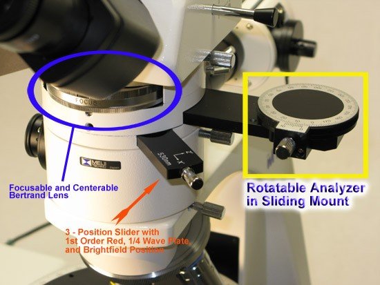
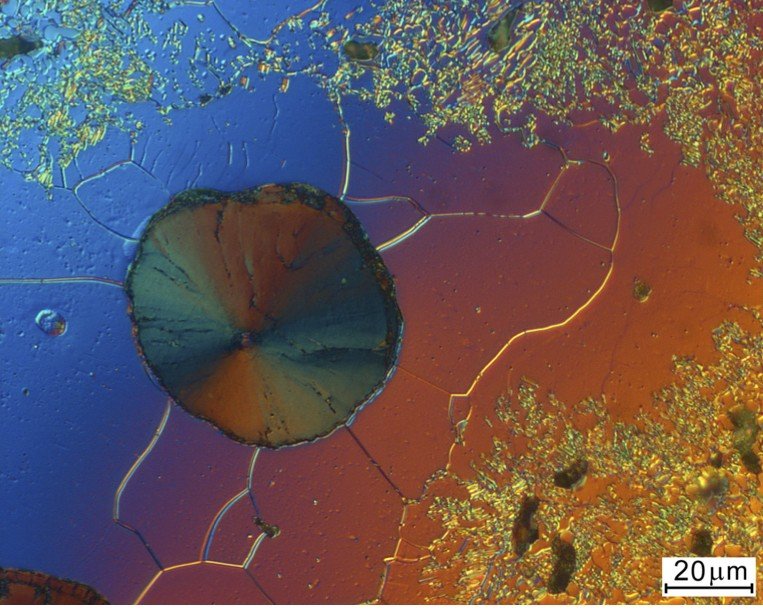
Polarized light microscopy is a useful tool for distinguishing between singly refracting (optically isotropic) and doubly refracting (optically anisotropic) material. Quantitative measurements of optical anisotropy are used in the optical analysis of doubly refracting or birefringent materials under polarized light. These measurements are made with the aid of accessory plates called compensators and retardation plates. Retardation plates have a fixed optical path difference and compensators have a variable optical path difference. The intermediate tube of BIOIMAGER polarized light microscope houses is either a sliding or rotatable analyzer, a sliding Bertrand lens and a slot for insertion of retardation plates and compensators.
Polarizing Microscope Types:
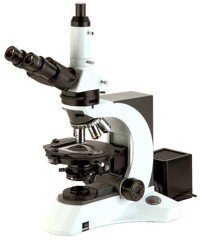
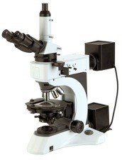
Polarising microscopes are offered withreflected (incident) light onlytransmitted light only (for transparent samples), or with both reflected & transmitted lights.
All polarising microscopes come with brightfield (BF) imaging and polarized imaging capability. There are some models that support brightfield and darkfield imaging.
Most of polarizing microscopes come in upright mode. If you need to have either an inverted microscope with polarization rather than upright, or need a microscope with all imaging capabilities (brightfield, darkfield, polarizing, DIC Nomarsky, or fluorescence), then please look at our inverted metallurgical microscopes such as BMI600 , or upright metallurgical microscopes such as BMU500DIC.
 |
 |
|
| reflected (incident) light only | transmitted light only | both reflected & transmitted lights |
All of BIOIMAGER polarizing microscopes include a polarizer, rotatable analyzer, Bertrand Lens, strain-free objective lenses and 360′ degrees rotatable stage which can be supplied with a mechanical XY stage to mount a 1×3″ slide or 2×3″ slide. Depending on the models, most polarising microscopes contain compensator (test plate), quartz wedge, halfwave
plate, quarter-wave plate, swing-out condenser, wide field eyepieces with reticle.
Basic Polarized Light Microscopy Terminology:
Polar: A device which produces plane polarized light from natural light.
Plane-Polarized Light: Light with only on vibration direction present.
Polarizer: A polar placed in the light path before the specimen.
Analyzer: A polar place in the light path after the specimen. The analyzer is removable from the light path and may be rotatable. The analyzer is used to determine the optical effects produced by the specimen either in plane or polarized light.
Strain Free: A term used to signify that microscope objectives and condenser lenses are selected to have a minimum amount of internal stress in the glass. Strain free optics offers little or no contribution to the optical path difference of the specimen.
Bertrand Lens: The Bertrand lens is located above the analyzer. the eyepiece and Bertrand lens act as a system to focus on the back image plane of the objective. The Bertrand lens’ main function is to view interference figures (conoscopic images) which appear in the back image plane of the objective when specimen is viewed between crossed polars using highly convergent light from the condenser.
Birefringence: A quantitative expression of the separation of a light beam as it penetrates a doubly refracting object into two diverging beams.
Conoscopic Figure: A pattern consisting of isogyres and/or isochromatic curves formed in the back image plane of the objective also referred to as an interference figure.
Conoscopic Observation: Observation of the back focal plane of a light microscope objective with a Bertrand lens or phase telescope, using a cone of light from the condenser.
PleoChroism: A property exhibited by certain crystals of absorbing selectively various wave lengths of light and displaying different colors when looked at in the directions of their various crystal axes.
APPLICATION NOTES:
 |
Application: High quality image capture with up to 1000x magnification by means of light-optical microscopes and high image resolution such as 12.5 mega pixels
Target: |
 |
Requirements: • high luminosity with polarized light and very good differential interference contrast with great luminous efficiency • high digital resolution of the images • a fast working process |
 |
Solution:
BPM800RT Microscope with ProgRes® C14, ProgRes® C14plus or BRC-1600 16MP Camera. Features: |
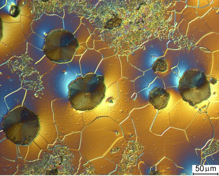
Samples Images
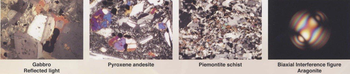
Images of Gabbro, Pyroxene andeite, Piemontite schiest, Biaxial interference figure Aragonite:
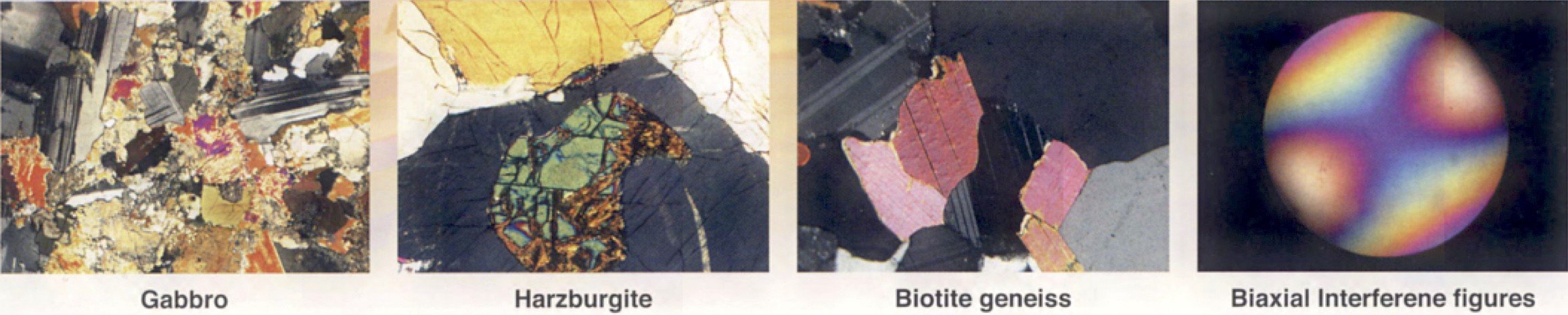
Images of Gabbro, Harzburgite, Biotite genesis and Biaxial interference figures:
Video Demonstration:
1. Introducing a Meiji Trechno (japan) MT9000 Polarizing Microscope
2. What is Polarized Light?
3. Principles of Polarising Microscopy