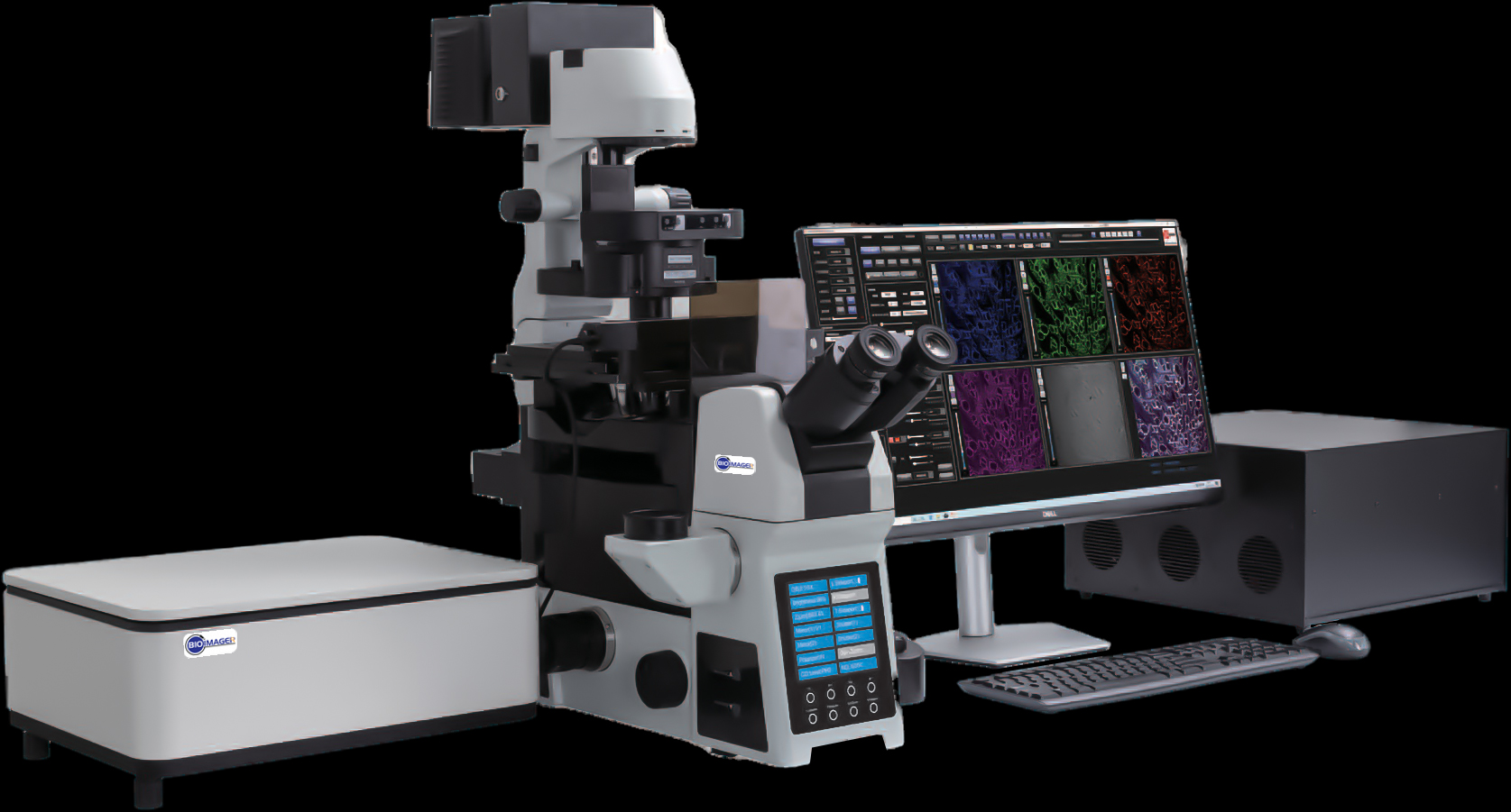
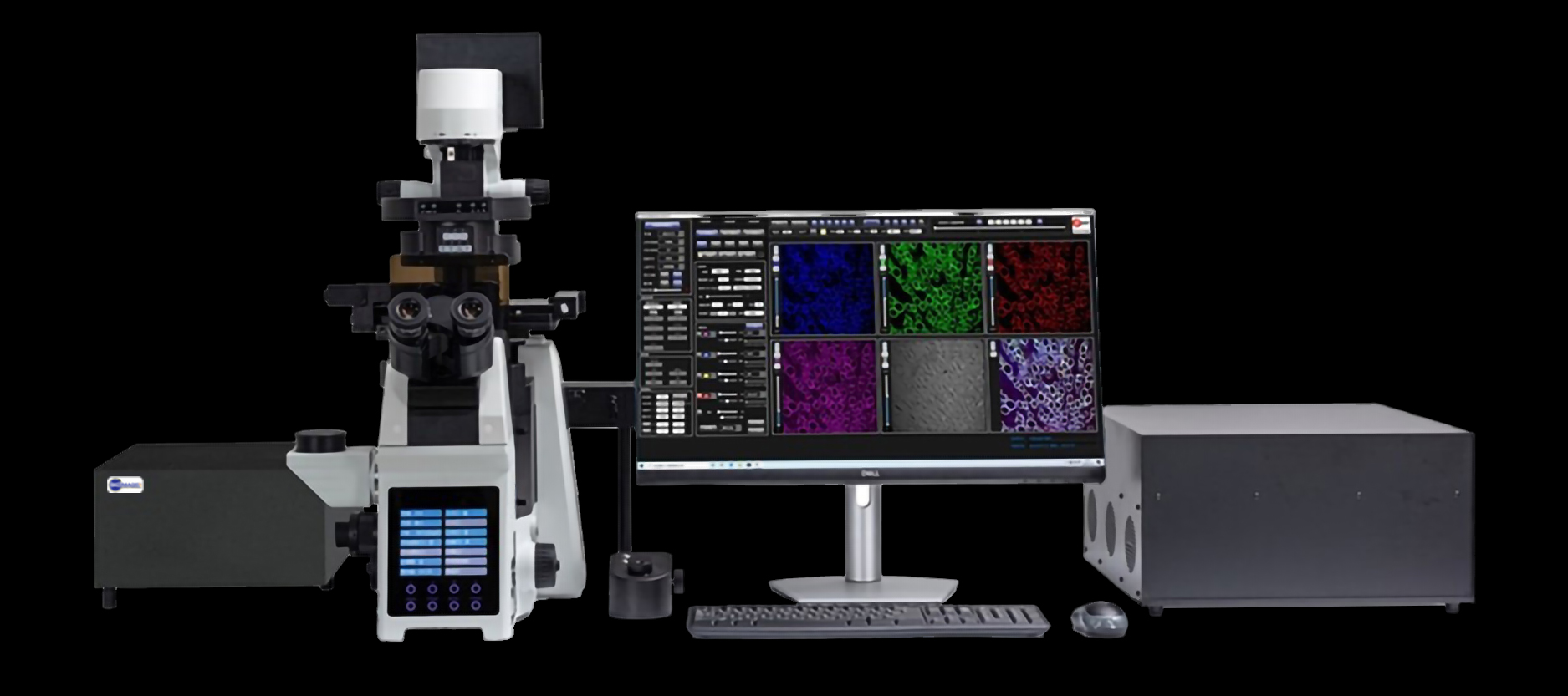
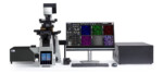
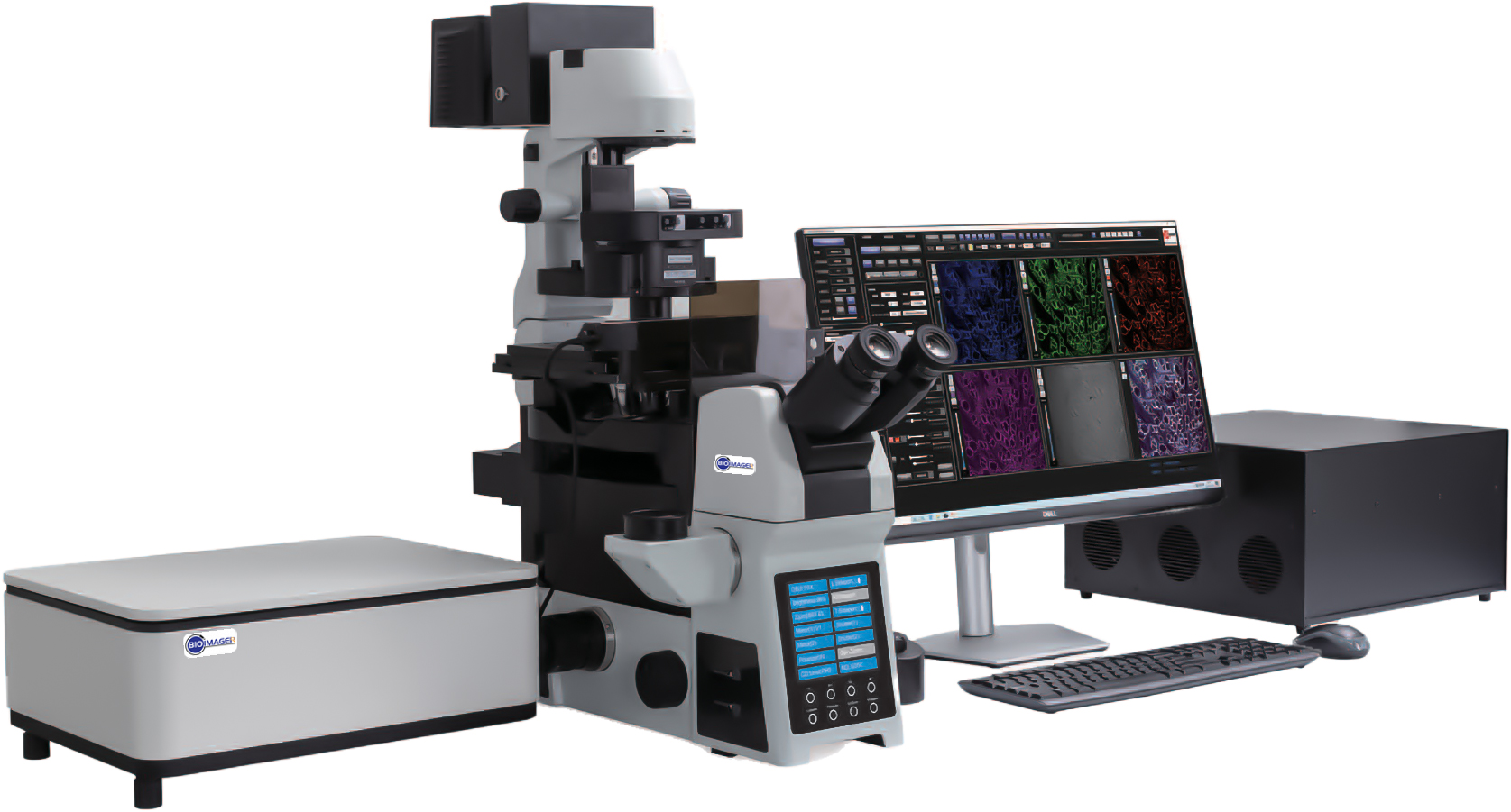
BCF-600 Laser Scanning Microscope – Advanced Imaging for Biomedical and Fluorescence Research
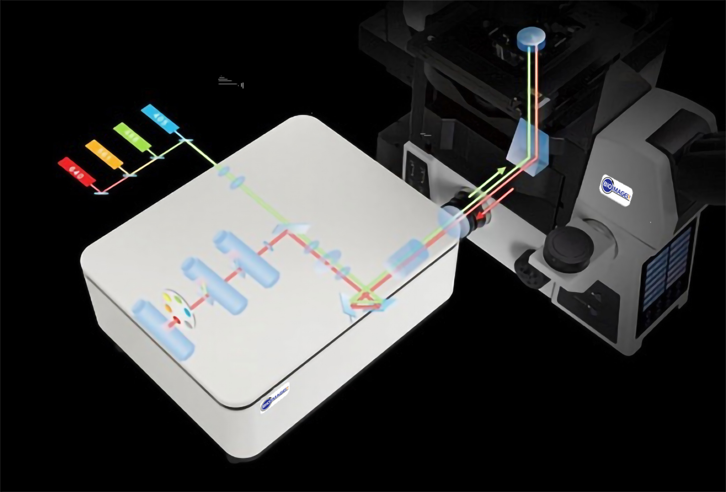
The BCF-600 Laser Scanning Confocal Microscope is designed for high-performance imaging in biomedical research, offering unparalleled precision and speed. Equipped with a versatile laser system featuring 405nm, 488nm, 561nm, and 640nm wavelengths, the BCF-600 is ideal for morphology, immunology, genetics, and other advanced research fields.
With adjustable laser control and polarization-maintaining single-mode fiber output, the microscope ensures precise, high-quality imaging. It supports high-speed X/Y dual-axis optical scanning using a galvanometer and delivers an extensive field of view (14mm x 14mm), with resolutions ranging from 512 x 512 to 4096 x 4096 pixels. The adjustable pixel time and fast scanning speeds offer flexibility and efficiency for a variety of research applications.
The BCF-600 also provides a range of customizable features, including pinhole options (φ30/40/50μm), a four-channel spectroscope, and a six-position electric turret filter system. Its GaAsP PMT probe offers enhanced sensitivity with up to 45% quantum efficiency at 500nm, enabling detailed fluorescence imaging.
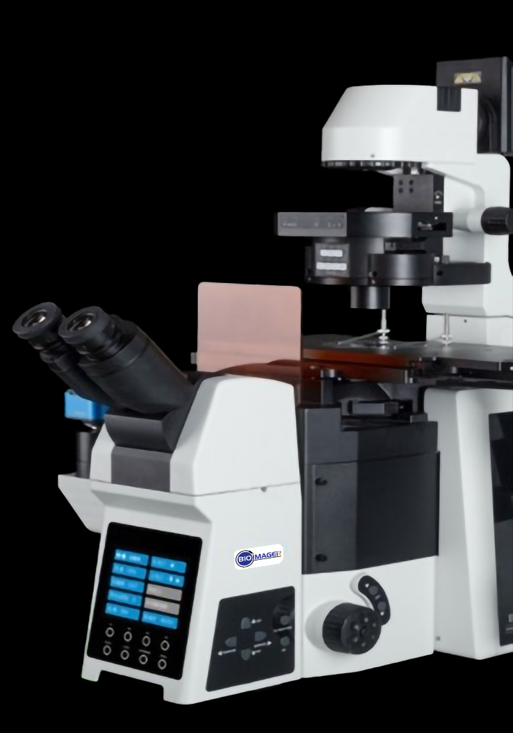
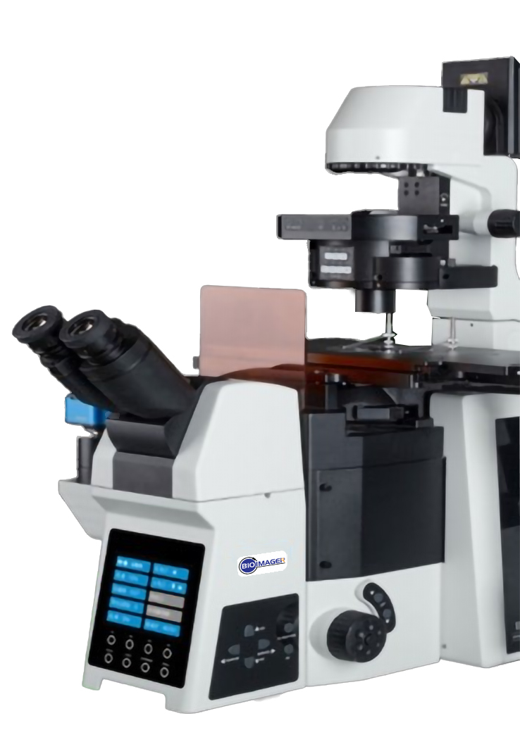
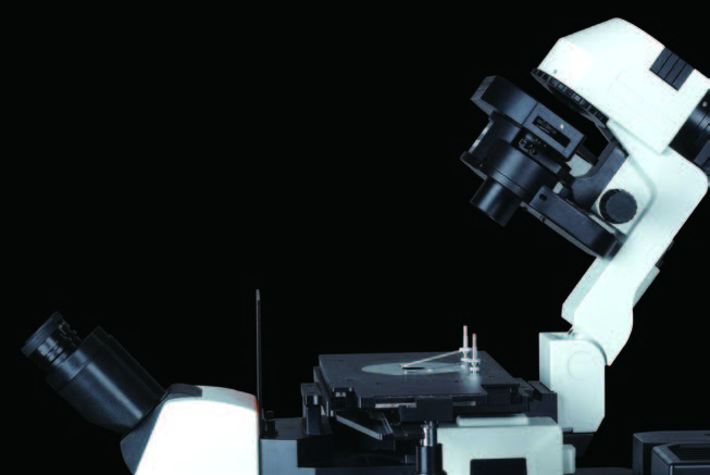
For user comfort and precision, the microscope includes a 20-45 degree tilting binocular head, high eyepoint wide-field eyepiece, and an infinity color correction optical system. The electric stage allows precise control, with a repositioning accuracy of ±1μm, making it adaptable to various sample sizes, including 35mm culture dishes.
The BCF-600 supports advanced scanning modes such as two-dimensional (XY), three-dimensional (XYZ), and four-dimensional (XYZT) time-lapse imaging. The eight-position fluorescent illumination system further enhances fluorescence experiments, offering automatic positioning and diaphragm control for precise illumination.
For researchers, the BCF-600 also provides advanced data management and processing features, including multi-color fluorescence co-localization, Z-stack analysis, and large image mosaics. Its integrated software offers seamless hardware connections, automatic storage, and autosave scanning parameters, ensuring that no data is lost during your experiments.
The height of electric Z-axis is able to be fast adjusted according to real-time image. Auto focusing by AF key, eliminate the step of fine tuning, improve work efficiency.
Integrated control buttons on both sides of the frame, can quickly switch or rotate the condenser, brightness, objective lens, attenuator disc and fluorescence disc, improve the operation convenience.
The tilting transmitted illumination
The transmission system adopts the tilting
structure to ensure Larger working space,
easier to change samples.
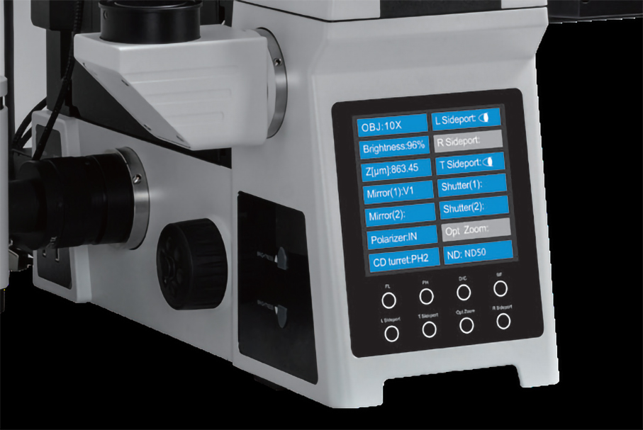
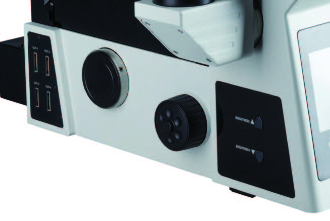
Front LCD panel
It is able to display the status of electric parts in real time, and set the observation mode, switch the light brake, etc., which greatly improves the user experience and makes the research work more convenient.
High scalable
The big frame provides sufficient space for third-party configurations. Single-layer optical path or double-layer optical path can be selected as required. It can be loaded into 16 filters at most, providing maximum scalability for the in-depth research.
Distinguished confocal imaging optical components
Innovative confocal pinhole unit
Pinhole design is based on the principle of light reversibility.The excitation light of the lamp and the emission light of the sample pass through the same pinhole, and they keep a 100% conjugate relationship. It not only ensures the acquisition efficiency of fluorescence signal, but also improves the filtering of non-focal plane signal, for the higher detection sensitivity and better image resolution
Controller probe unit
The probe unit consists of a 6-position electric filter turntable with 4 filters as standard and a single high-sensitivity multi-base photomultiplier tube (MA PMT, QE ≥ 25%@500nm), is able to easily and quickly automatically complete multicolor fluorescence confocal imaging.
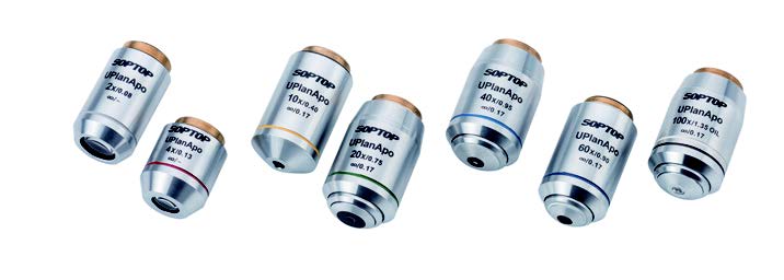
APO series apochromatic objectives
Converging the optic axises of red, green and blue to one focal plane, correcting the axial chromatic aberration of violet light, the original color of samples is able to be presented. And the resolution and effective magnification are improved based on large numerical aperture.
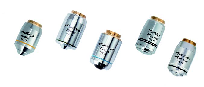
SAPO series super apochromatic objectives With large numerical aperture, excellent color difference correction and flat field, more uniform, bright and high-resolution fluorescence images can be obtained.
Confocal software
Support single channel or multi-channel 2D Imaging (XY), 3D imaging (XYZ), 4D imaging (XYZT) and multi-site scanning. It is available for imaging, photobleaching and photostimulation within a user-defined ROI, as well as Z-Stack imaging, jigsaw puzzles, scale correction, filtering processing, data recording, etc.
BCF-600 Laser Scanning Confocal Microscope – High-Precision Imaging for Advanced Biomedical Research
The BCF-600 Laser Scanning Confocal Microscope offers high-precision observation and fast, accurate analysis, making it the ideal solution for a wide range of applications in morphology, physiology, immunology, and genetics. This advanced microscope is designed for cutting-edge biomedical research, delivering superior imaging results with speed and reliability. Whether you’re working on genetic analysis, cell structure observation, or immune response studies, the BCF-600 provides exceptional performance and precision, enhancing your research capabilities.
BCF-600 biological application:
Cell imaging
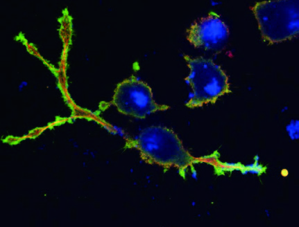
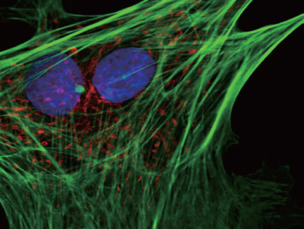
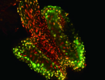
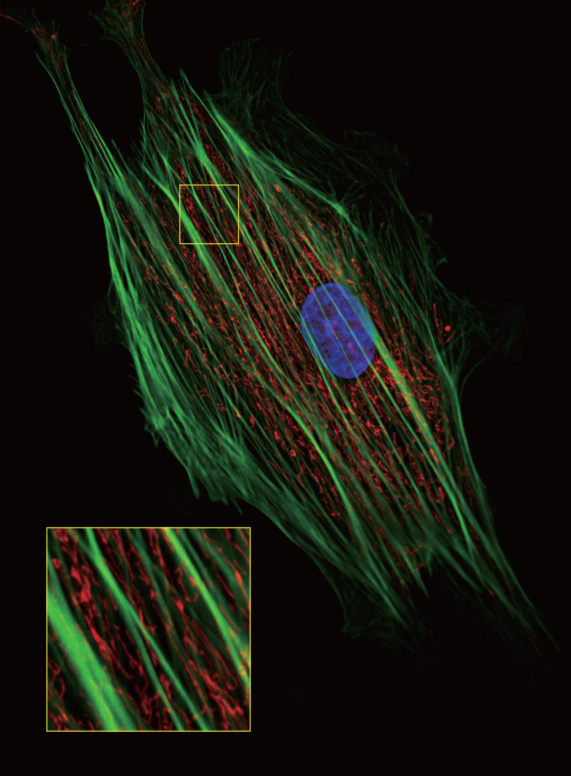
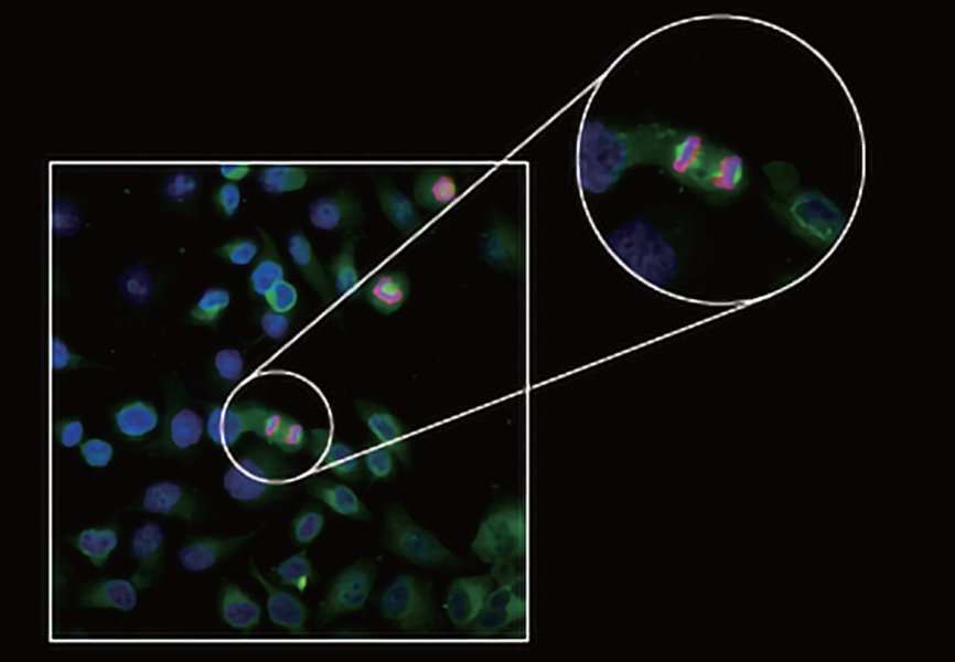
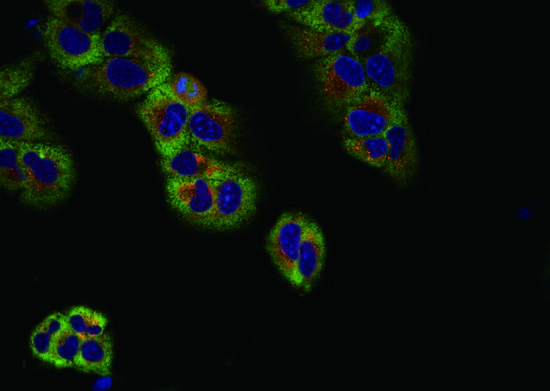
BCF-600 is able to accurately image all cells labeled with various fluorescent proteins and multicolor probes, studying the fluorescence colocalization, dynamic properties and spatial relationships of two or more target proteins. Besides, CLSM600 can achieve the morphological structure of 3D cell culture such as organoids/globules by 3D reconstruction, finding out more hidden information.
HELA Cell (Peking University Health Center) 60X
Human hepatocytes (Zhejiang University) 40X
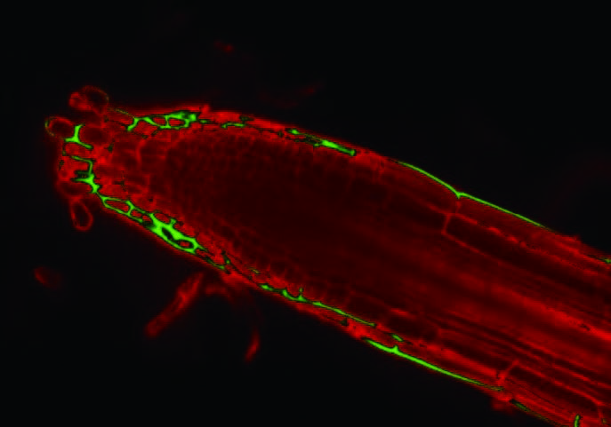
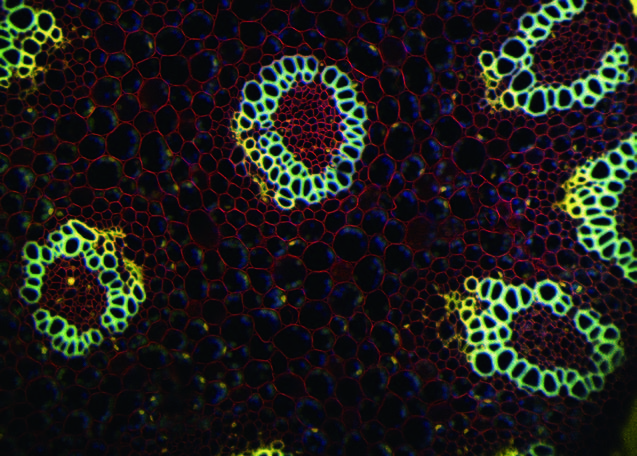
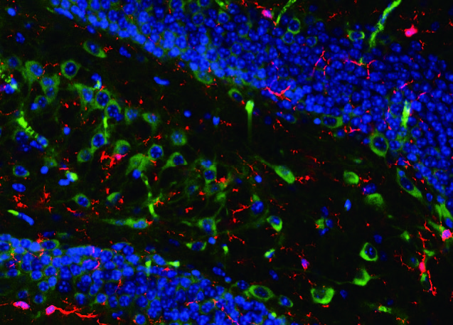
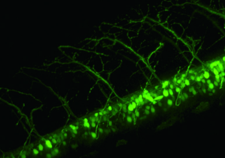
Histopathological sections of animals and plants
The layer scanning of BCF-600 is suitable for different histopathological sections of animals and plants, especially for large tissue. Much more details and more accurate data are available.
Convallaria rhizome (Zhejiang University) 20X
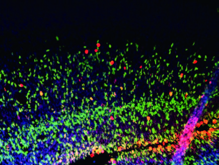
Hippocampal slice (Chinese Military Academy of Science) 40X
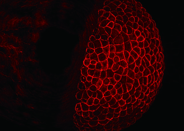
Viviperception of model animals and plants
In general, common model organisms such as zebrafish, fruit flies, nematodes, and Arabidopsis thaliana are characterized by large size, complex structure, and high density. With wide FOV and layer scanning, BCF-600 is an ideal tool to get fine structure image and details at different depths.
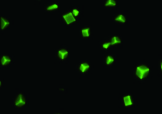
In the field of biomphotonics, the research heat of biological information and nanomaterials is increasing. In the study of photoelectric materials, BCF-600 can cooperate with the living cell environment monitoring module to observe the process of interaction between materials and cells for the fusion of new functional materials, inorganic nano hybrid materials and living cells.
Functional materials (Wuhan University of Technology) 60X
Laser light
Laser
405nm/50mW、 488nm/50mW、 561nm/50mW、 640nm/40mW
Laser control
The switch and intensity are adjustable
Output mode
Polarization-maintaining single-mode fiber
Scan module
X/Y dual axis high speed optical scanning galvanometer
Filed of view: 14mm X 14mm ( ≥ 19)
Scan unit
Scanning element: 512 X 512 ~ 4096 X 4096
Pixel time: 0.5μs ~8μs
Standard scanning speed: 1fps (512 X 512, 2μs), fast scanning speed: 3fps (512 X 512, 0.5μs)
Zoom scanning: 1X~50X
Pinhole
φ30/40/50μm Option
Spectroscope & filter
Four-channel spectroscope: 405/488/561/640nm
Six-position electric turret with 4 standard filters: 445nm/40, 530nm/43, 607nm/36 and 685nm/40
High eyepoint wide filed plan eyepiece PL10X/22mm, diopter adjustable, micrometer attachable
Objective
Infinity plan apochromatic objectives
Infinity plan super apochromatic objectives
Frame
Low position coarse and fine coaxial electric focusing mechanism, range 10.5mm, precision 1μm; built-in electric upper camera port, built-in electric left camera port, dual optical paths; with fluorescent light barrier; 6-position electric nosepiece with DIC slot and C-mount adapter
Stage
Manual mechanical stage, size: 300mm(X) x 240mm(Y), moving range: 135mm(X) x 85mm(Y)
Electric stage, size: 260mm(X) x 153mm(Y), moving range: 110mm(X) x 75mm(Y), with operating handle; Max. speed 3mm/s, repositioning accuracy ±1μm, 35mm culture dish adaptable
Condenser
Electric sever-position condenser, NA.0.55, WD27mm; 3 positions for φ30mm (phase contrast), 4 positions for φ38mm (DIC); support for bright filed, phase contrast and DIC (including polarizing kit)
Fluorescent illumination
Eight-position fluorescent illumination system with automatic positioning, with electric diaphragm for fluorescent light, with fluorescent filters B/G/UV etc.
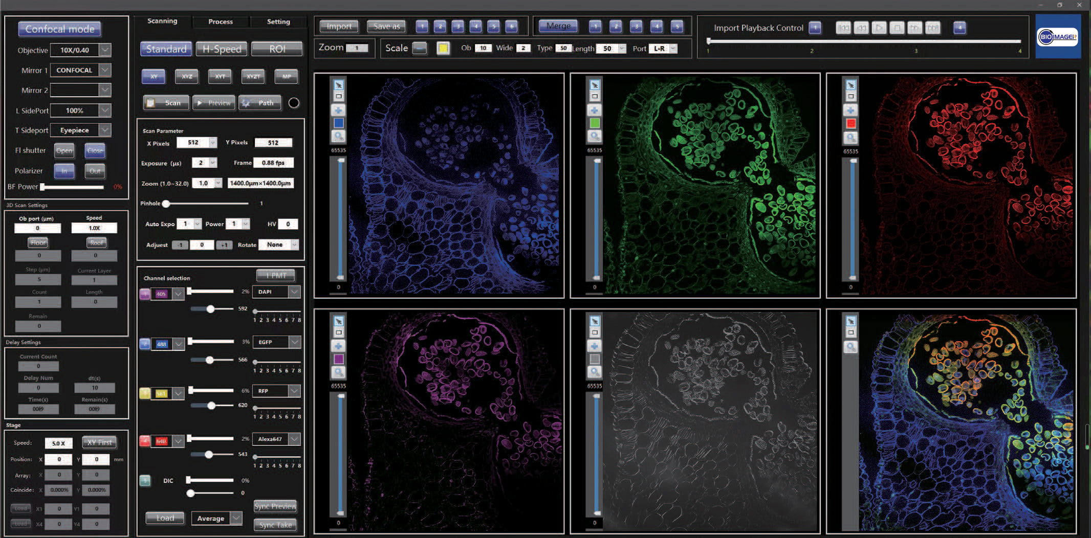
Operating software
Scanning image
Photo and preview parameter self-adaptive; two rescan processing; image rotation output; full vision and ROI scanning image; monochromatic or polychromatic two-dimensional image (XY), three-dimensional image (XYZ), four-dimensional image (XYZT) and multi-point scanning
Process
Multicolor fluorescence co-localization, fluorescence and DIC image addition; correcting reticle; filtering processing; Z-Stacke analysis and large image mosaic
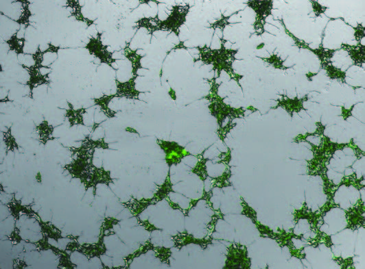
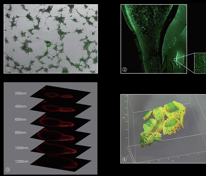
①Hek 293t cell 60X DIC
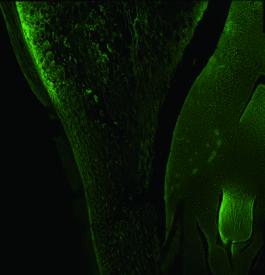
②Corn root 40X Large image mosaic
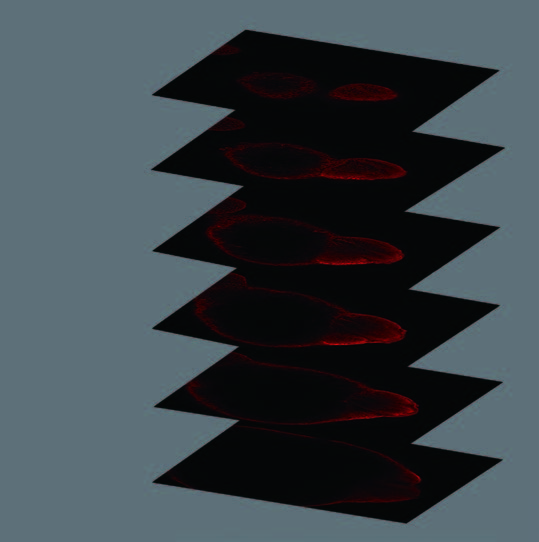
③Zebrafish 20X Z-Stack
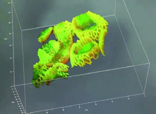
④Organoid 3D reconstruction
Operating software
Scanning image
Photo and preview parameter self-adaptive; two rescan processing; image rotation output; full vision and ROI scanning image; monochromatic or polychromatic two-dimensional image (XY), three-dimensional image (XYZ), four-dimensional image (XYZT) and multi-point scanning
Process
Multicolor fluorescence co-localization, fluorescence and DIC image addition; correcting reticle; filtering processing; Z-Stacke analysis and large image mosaic


Reviews
There are no reviews yet.