
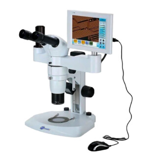
MS-6 LCD Industrial Stereo Zoom Microscope
Introduction
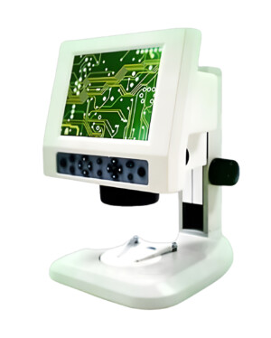
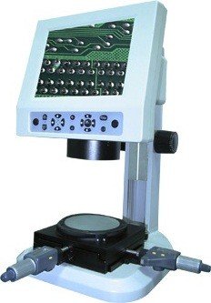
The digital LCD biological microscopes integrate traditional optics and digital technology.It combines the tablet pc on microscope to take the place of the binocular head. A digital camera is built in the head of the microscope, users can observe image on LCD screen. These microscopes make the observation more comfortable and resolve the fatigue caused by using a traditional microscope for a long time. These microscopes integrate magnification, digital enlargement, imaging display, capture photo and video and store captured photos and videos. Users can transfer images and videos to PC via included USB cable and internet.
Specifications
Digital Parts:
| Photoreceptor Chip | 3.0MP 1/2” Digital Photoreceptor Chip |
| Resolution | 2048×1536 |
| Frame Rate | 12fps @ 2048×1536
30 fps (MAX) |
| Data Interface | USB2.0 (High Speed) |
| Signal-to-noise Ratio | > 45 db |
| Imaging Mode | Planar array |
| Scanning Mode | Progressive Scanning |
| Exposure | Manual, Automatic |
| White Balance | Manual, Automatic |
| Wireless network card | 802.11n with wireless transmission rate: 300M (Max) |
| Network Card | Wired Ethernet Interface |
| Audio | One Microphone Input, One Audio Output |
| Video | One VGA Output |
| USB Interface | Five USB Interface (High Speed) for Keyboard, Mouse & USB Storage |
| Screen | 13.3″ 16:9 32-bit LCD Screen |
Software Parts:
| Preview Resolution | 2048*1536, 1600*1200, 1280*1024, 800*600 |
| Collection Resolution | 2048*1536, 1600*1200, 1280*1024, 1024*768 |
| Automatic Exposure | Adjustable the exposure parameters, brightness of the field of view, target value. |
| Manual Exposure | Adjusting the exposure time & gain parameter |
| Automatic White Balance | Reproduce the real-time image automatically. |
| Manual White Balance | Adjusting RGB, Gamma, Contrast, Saturation and etc |
| Full-screen View
Regional Preview |
Automatic Adjustment for the 3.0 MP Dynamic images to adapt to the resolution of the screen. Selected parts can be amplified for interests & details. |
| Static Images Taking | Single-frame capturing on the dynamic images, and storage in the disk by automatic or manual |
| Timing Images Taking | Capturing according to the time interval. The frames and time interval can be adjusted according to the users’ requirement. |
| Dynamic Images Recording | Capturing the dynamic images shown on the microscopes, and numbering them automatic or manual, and store these images in the disk. |
| Compression Dynamic Images Recording | Record the dynamic images in MPEG4. |
| Geometry Size Testing | Geometry Parameter: Line, curve, round, rectangular, angle, size, perimeterUnits: MM, CM, Inch and etc |
| Calibration & Scale | |
| Images Transformation & Geometry Correction | Level, Vertical, 90° (counterclockwise), 90° (clockwise), any angle rotatable |
| Tools for Region Selected | Rectangular, round and arbitrary shape |
| Function of magnifier | Reflect the details of the images much sharper |
| Image Processing | Hue processing: negative image, gray processing, hue adjustment, Brightness, contrast, and RGB.Image Enhanced: Histogram Balance and filter
Others: edge enhanced, edge testing, mosaic, sharpen, confusing, soften, exposure and etc. Special processing: relief, diffuse, perspective and etc. |
| Fluorescence Composing | |
| Printing | Reproduce the real size of the image |
| Marking | Mark on the images, (scale, character, and images) |
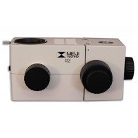
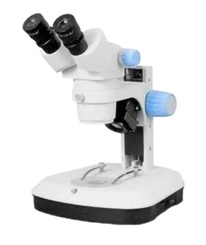
Optical parts
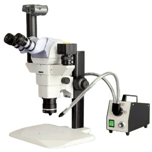
Viewing Head: Binocular head inclined at 45°-360° rotatable, adjustable interpupilary
Distance (52-76 mm),
Magnification: 0.8X-5X
Zoom Ratio: 1:6.3
Eyepiece: WF10X/22
Illumination: Reflected & Transmitted Illumination
Reviews
There are no reviews yet.