


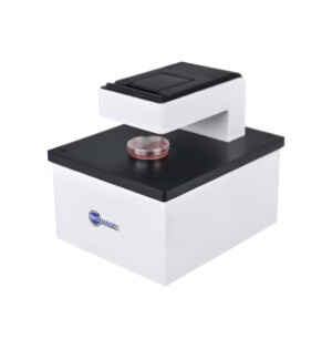
What is an IncuScope?
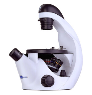
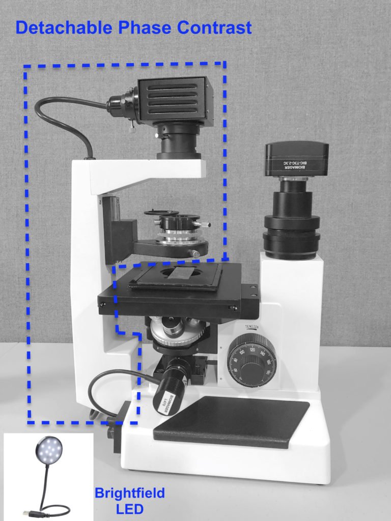
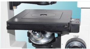
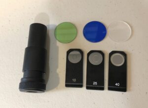
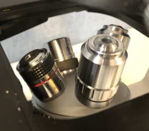
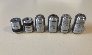
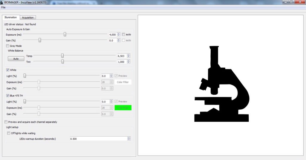
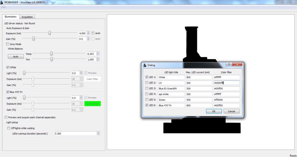
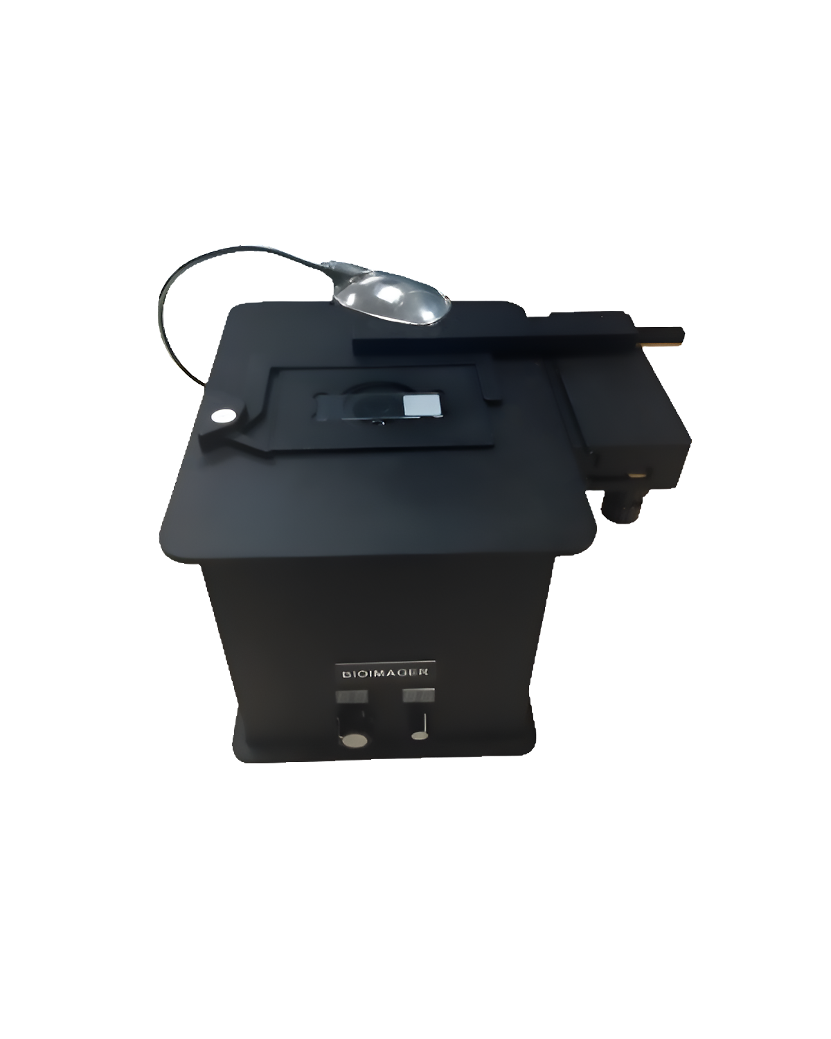
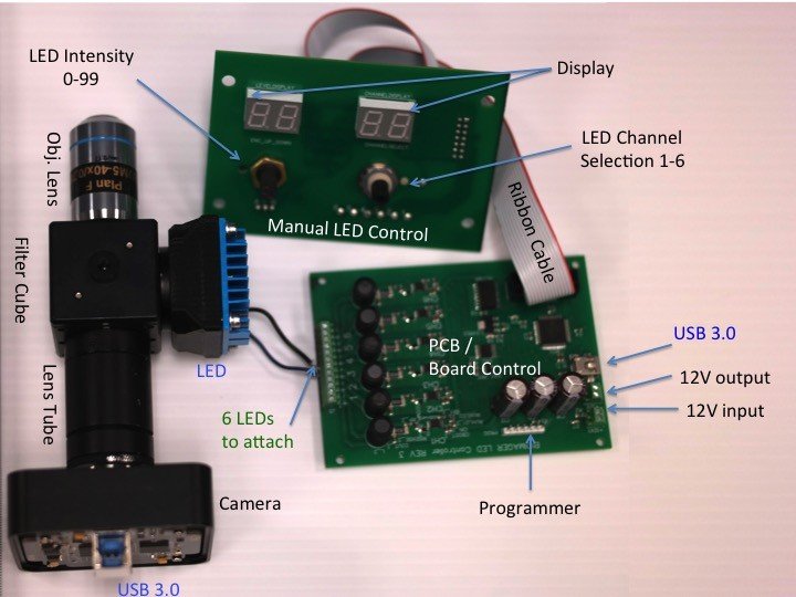
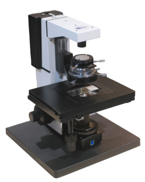
IncuScope is a compact inverted fluorescence microscope that fits on a shelf inside an incubator or inside a hood. It has brightfield, phase contrast and fluorescence imaging capability with optional darkfield imaging. It can be either used with camera only or with a trinocular head attached to a camera. Thus it will be a stand-alone microscope to use on the bench or keep inside an incubator for video time-lapse imaging. This system can run on a laptop or desktop using USB 3.0 or USB 2.0 cable, allows capturing images, and controlling the LEDs. It has several options of objective lenses (from 2x to 150x) and fluorescent filters (from UV to IR). The IncuScope is the most simple and affordable live cell imaging device in the market.
 |
 |
 |
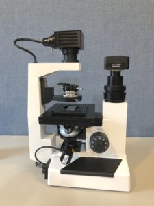
| On the bench
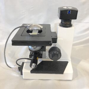
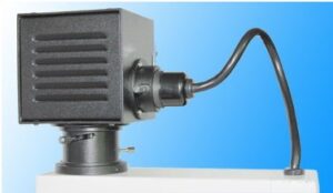
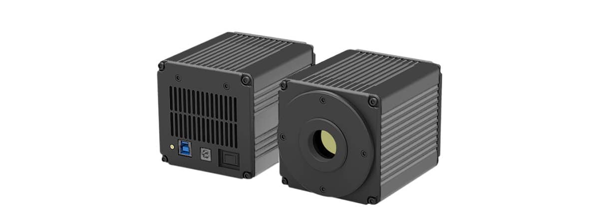
Brightfield & Fluorescence camera attachment |
Inside a hood
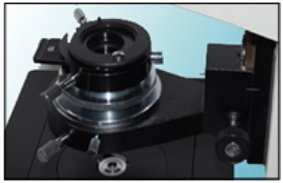
Phase Contrast & Fluorescence camera attachment |
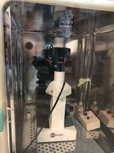
Inside an incubator
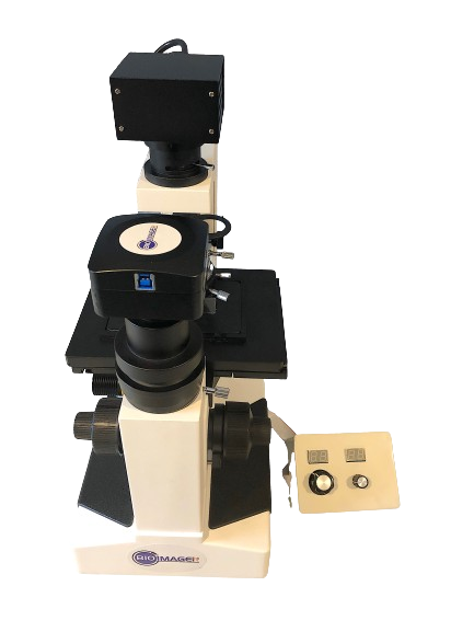
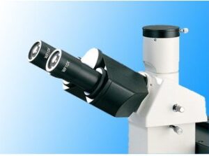
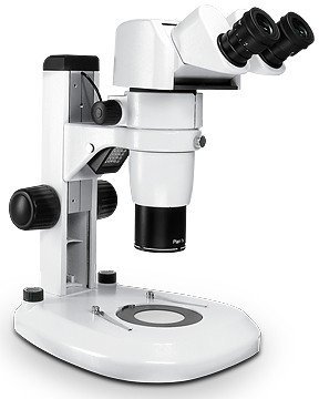
Fluorescence & Phase Contrast trinocular head & camera |
Applications
When you need to have a complete access of a biological microscope inside an incubator, IncuScope is a perfect choice. If you need to monitor your cells under microscope in hypoxy conditions and do not like to spend $25,000 for a miniature incubator / heating stage to add to your microscope, IncuScope IS250 will do the job you need. Some examples of IncuScope Applications are (but not limited to):
- Cell Proliferation and confluency study
- Cell growth, death and differentiation kinetics
- Cell Counting
- Cell Migration and wound healing
- Cell Cycle
- Stem cells development
- Cancer Cells
- Microfluidics study
- Hypoxy Condition
Why IncuScope IS250? Why is it better than the other similar products?
The IncuScope IS250 series has several advantages over the competitors such as:
- Price: it has the lowest price of the incubator microscope in the market while its image quality and performance stay superior. For instance:
- Lumascope 560 series with BF and FL imaging, single obj lens costs $7,500 USD, plus XY stage for $750 and each sample holder at $130. If you need phase contrast, add another $3,350 for single obj lens.
- EVOS, with similar package starts at $18,000
- Juli with similar package starts at$ 25,000
- Image quality talks. You compare it to believe.
- Flexibility in design: This incubator microscope has the most diverse flexible design. You can select any combination of camera and c-mount adapter, and any fluorescence filter design.
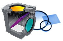
- Unique Feature: darkfield, polarization imaging is possible. The other incubator products can not offer this.
| IncuScope
250 |
LumaScope
560Ph |
EVOS
M5000 |
|
| Viewing Head | Trinocular included | Not Possible | Not available |
| Objective Lens | Multiple (4 or 5) | Single | Multiple (5) |
| Imaging Capability | BF, Ph, FL
optional Darkfield & Polarizing |
BF, Ph, FL | BF, FL |
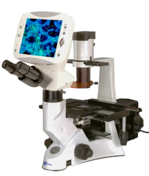
| Monitor | Optional HD Retina Display | Not possible | Available |
| Camera Selection | Several options of sensor sizes and speed
(or use your own) |
Fixed | Fixed |
| C-Mount adapter | Select from 0.37x to 1.2x | Fixed | Fixed |
| LED Selection | Customizable from 400nm to 1500nm, over 40 filter set combination | GFP only | Configurable |
| Dimension
(Depth x Width x Height) |
311 x 200 x 428 mm
12.2 x 7.9 x 16.8 in |
With XY Stage
240 x 267 x 378 mm 9.4 x 10.5 x 14.9 in |
460 x 330 x 360 mm
18 x 14 x 13 in |
| Weight | 4.5kg / 10lb | 4.5kg / 10lb | 16kg /35lb |
| Price | $7,495 | Starts at $10,850 | > $18,000 |
This video shows the video time-lapse of T Cells cultured in a 96-well plate and captured by IncuScope IS251 inside an incubator overnight (for 12 hours) using 25x Phase Contrast.Thanks Jamie McNicol at McMaster University, Hamilton, ON, Canada.
https://youtu.be/XOAXbv55iCQ


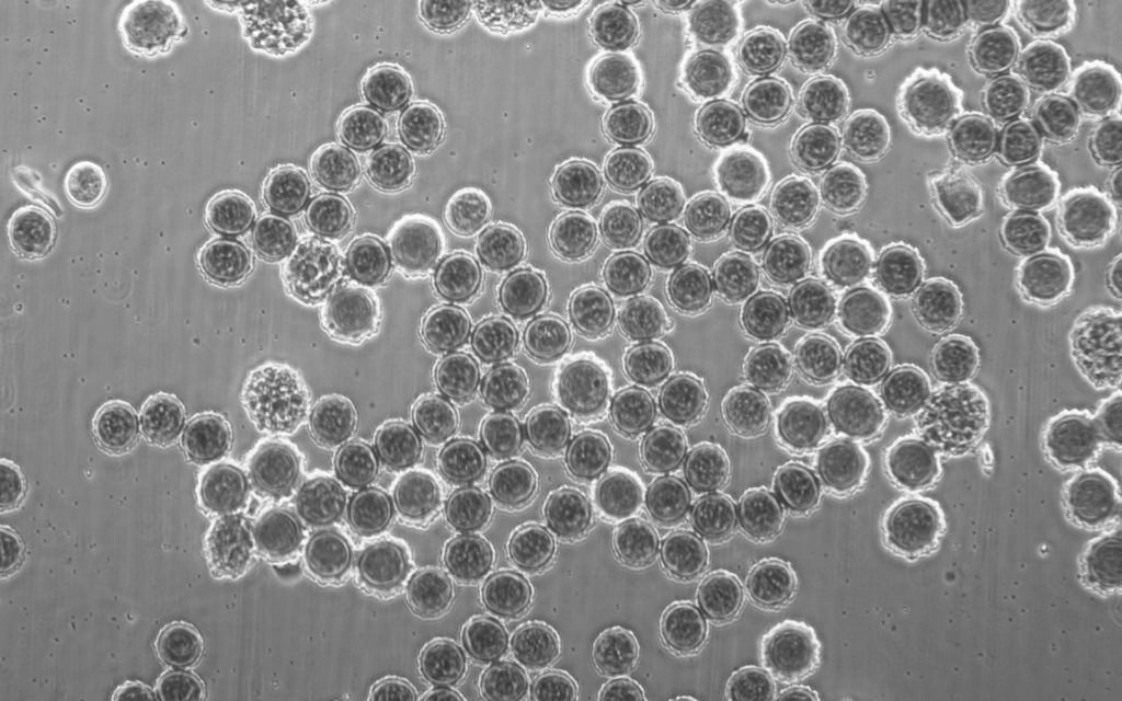 10x Phase Contrast
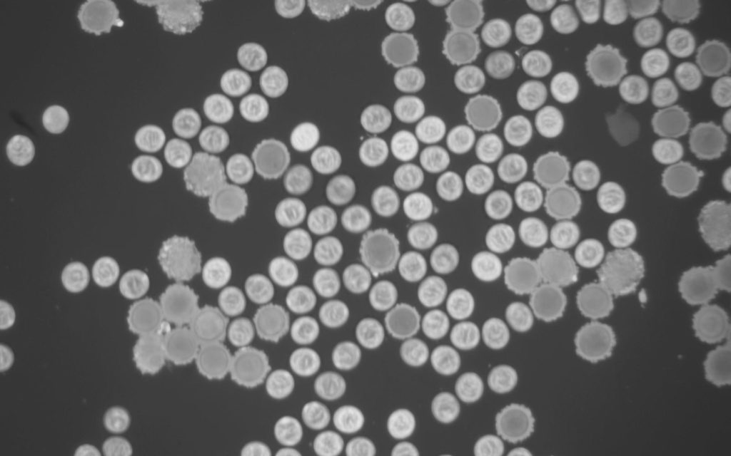 10x Fluorescence, Monochrome
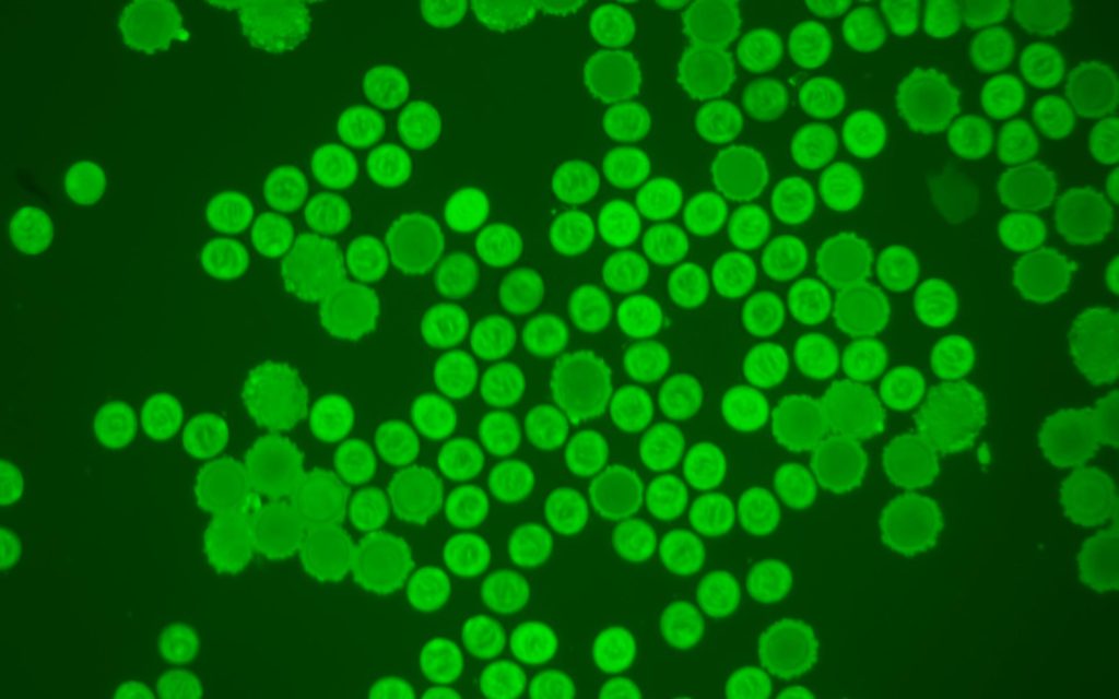 10x Fluorescence, labeled green (pseudo colour)
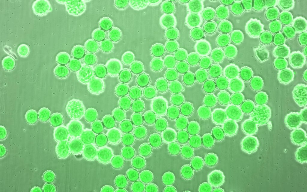 Merged / Superimposed image of phase contrast and fluorescence
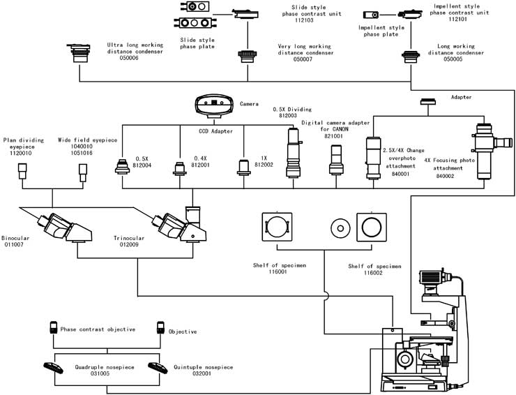
Reviews
There are no reviews yet.