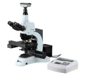
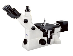
This microscope is a high level microscope which is typically designed for laboratory study. It adopts an Infinite optical system, reasonable structure and ergonomic design. With an innovative optical and structure design idea, excellent optical performance and easy to operate system, this laboratory biological microscope makes your laboratory works enjoyable.
Applications
This microscope can be used in institutes and laboratories to observe and identify the structure of various metal and alloy, they also can be used in electronics, chemical and instrumentation industry to observe the opaque material and transparent material, such as metal, ceramics, integrated circuits, electronic chips, printed circuit boards, LCD panels, film, powder, toner, wire, fibers, plated coatings, and other non-metallic materials and so on.
Main Features:
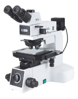
Four motor-drives: Objective nosepiece, XY stage and Z focus, controlled manually and automatically
Advanced technological digital dimmer with 100 steps of halogen lamp brightness division
High precision positioning leading new stage,
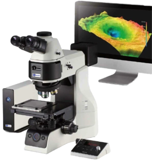
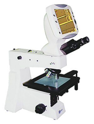
The clearest images with immediate access just by click on auto-focus, auto-scanning and auto-recognition features
Built-in dynamic focus mechanism; achieve the laboratory microscope automation through network remote control system,
widely used in medical diagnosis, pathological analysis and real-time teaching, etc.
3.2MP Camera with great Imaging software is included
With Infinite Optical System, Kohler Illumination, providing excellent optical quality and making Image perfect.
With low and forward position of C&F coaxial focus system, focusing knobs and brightness adjustable knob within reaching location, stable integral structure, and reasonable ergonomic design, making operators feel more comfortable and effective.
Four motor-drives: Objective nosepiece, XY stage and Z focus, controlled manually and automatically
Advanced technological digital dimmer with 100 steps of halogen lamp brightness division
High precision positioning leading new stage,
The clearest images with immediate access just by click on auto-focus, auto-scanning and auto-recognition features
Built-in dynamic focus mechanism; achieve the laboratory microscope automation through network remote control system,
widely used in medical diagnosis, pathological analysis and real-time teaching, etc.
3.2MP Camera with great Imaging software is included
With Infinite Optical System, Kohler Illumination, providing excellent optical quality and making Image perfect.
With low and forward position of C&F coaxial focus system, focusing knobs and brightness adjustable knob within reaching location, stable integral structure, and reasonable ergonomic design, making operators feel more comfortable and effective.
Microscope Specifications
Models of BMU500A:
RF
TRF
Optical System
Infinite optical system
●
●
Viewing Head
Siedentopf trinocular viewing head, inclined at 30°, interpupillarty distance 48mm-75mm
●
●
Eyepiece
Extral wide field eyepiece EW10×/22, eyepiece tube Φ30mm
Exposure: Manual/Auto Exposure Process, Exposure Time; Adjustable: 1~500 ms
SNR: 43dB
Dynamic Range: 61dB
Connecting Mode: Insert it into Eyepiece Tube of Microscope Directly or Using Standard C Mount
Image Output: USB2.0, Directshow & Twain
Working Temperature: 0°C ~ +60°C
System Windows: 2000/XP/Vista/Win7 32bit or 64bit
With High Resolution chip (3.0M), Providing High Quality Image and Widely Used in Academic and Medical Field for High Precision and Resolution Image Capturing and Processing; By USB2.0, getting Real-time and Non-compressing Video Data and Capturing Image Directly;
Video 1: Main Body and Performance Overview:
Video 2: Imaging Capability: Brightfield imaging with transmitted or Reflected Light
Video 3. Fluorescence Imaging
This video demonstrates Bioimager BUM360FLED-A LED Fluorescence attachment to use with a standard upright microscope such as BMU500A.. It comes with 5W LED of Violet (V), Ultraviolet (UV), Blue (B) and Green (G) LEDs, and its correspondent excitation and emission filters. The video demonstrates the optics and mechanics as well as sample images at different magnifications.
Software
High resolution image processing software is especially designed for digital microscope; it has friendly operation interface, stable performance and powerful function, very easy to operate. And can be widely used in various optical micro fields, such as teaching, researching, electronic checking and so on.
Software Features
ScopeImage 9.0 is image-processing software professionally designed for digital microscope. It allows you to view, capture, edit, record, zoom, measure, and process microscope images. Specific features include as follows:
Multi-language Support: English, Germen, French, Japanese, Chinese and Arabic; It can switch easily between the languages.
Preview modes: active image, freezing, fit to window, actual pixels and full screen preview, all in a user-friendly environment.
Convenient color adjustment of the active image, containing brightness, contrast, saturation, RGB value, white balance and so on.
Special design color database; Users can associate the parameters of the color to the database, store or delete the color scheme.
Various image measurement tools include line, angle, rectangle, polygon and circle, move, delete or set the color of the objects, add text on the image, burn all the objects into the image for further use.
Automatic measurement and real-time measurement result display.
Basic image operations like other similar software are included in the Image menu. User-friendly image manipulation and enhancement.
Image flip function include horizontal and vertical flips of the active image, and rotate 90 degree, 180 degree, 270 degree or any angle degree of the captured image.
Image Zoom: Contain image zoom in, zoom out, 1:1 display.
System Requirements
Operating System: Windows 2000/XP/Vista/Win7 32bit or 64bit
DirectX 9.0 or higher (for Windows 2000)
Video adapter supports 24bit color or more, and 1280×1024 or 1024×768 resolution
CPU with 1.8GHz or more
System Memory 512MB or more
Display Memory 256MB or more
USB2.0 interface
Hard Disk Space 1GB for installation plus additional space for captured images.
Custom Microscopy package of this model was provided to several customers. These are some examples:
Montana State University, Bozeman, MT, USA
POSTECH University, S. Korea
International Sci. & Technol. Center (ISTC) Astana, Kazakhstan & Yerevan, Armenia
Reviews
There are no reviews yet.
Only logged in customers who have purchased this product may leave a review.


Reviews
There are no reviews yet.