Software
Showing 1–9 of 13 results
-
$USD 499.69 Add to cart
-
-
$USD 1,495.00 Add to cart
-
-
$USD 1,199.00 Add to cart
-
-
$USD 5,125.00 Add to cart
-
-
Showing 1–9 of 13 results
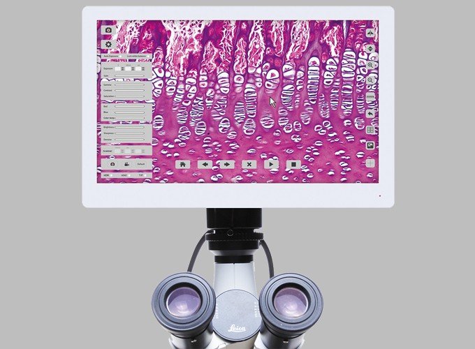
Microscopy imaging software
Microscopy imaging software is a type of software that is designed to help users acquire and analyze images and data from microscopes. There are many different types of microscopy imaging software available, each with its own set of features and capabilities. Some of the key features of microscopy imaging software include:
- Image acquisition: The software allows users to acquire high-quality images and videos from the microscope. This includes features such as adjusting exposure time, contrast, and brightness.
- Image analysis: The software enables users to analyze and measure the acquired images. This can include tasks such as counting cells, measuring distances and areas, and tracking movement.
- Image processing: The software can enhance the quality of images by reducing noise, correcting color balance, and adjusting contrast.
- Image management: The software provides a way to organize, store, and retrieve images and data. This includes features such as creating annotations and adding metadata to images.
- Integration with other tools: The software can be integrated with other tools, such as data analysis software and microscope hardware, to provide a complete microscopy solution.
Some popular microscopy imaging software packages include ImageJ, FIJI, Zen, and MicroManager. The choice of software often depends on the specific needs of the user and the type of microscope being used. Overall, microscopy imaging software plays a critical role in microscopy research by enabling users to acquire, analyze, and manage high-quality images and data.
Image Analysis in Life Sciences (Pathology, Immuno-histochemistry & Fluorescence) & Industrial Sciences (Quality Control, Physical Science & Particle Analysis).
| Life Science Image Analysis
These are just a few of the many life science image analysis applications our software supports:
|
Industrial Image Analysis
These are just a few of the many industrial science image analysis applications our software supports:
|
Life Science Image Analysis
Whether you are involved in drug discovery, cell biology, fisheries science or other life science research, if your work depends on images and the information they contain, we have the tools to make your projects a success.
These are just a few of the many life science applications our software supports:
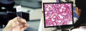
Pathology and Immunohistochemistry Image Analysis
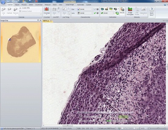
Image-Pro Premier software provides research pathologists with a vast range of tools for acquiring images, opening multi-resolution files, and identifying, classifying and quantifying nuclei, membranes and areas of stained or labeled tissue.
Pathology and Immunohistochemistry Analysis with Image-Pro Premier Software
Digital Pathology – from Morphology to Markers
Digital pathology encompasses a complementary range of advanced technologies optimized to facilitate the acquisition, organization, and utilization of information associated with biological cells, tissues, and organs. The ongoing evolution of specialized digital imaging and analysis tools allows pathologists to streamline their workflow in ever more intelligent ways, thus maximizing lab efficiency and productivity. Expert interpretation of both the qualitative and quantitative information obtained via these largely automated digital processes can effectively refine and expedite patient treatment.
Open Very Large Multi-resolution Images Acquired from a Slide Scanner
The Very Large Image tool in Image-Pro Premier allows pathologists to open large, multi-resolution files which were acquired using a slide scanner. Currently supported file formats include Aperio .svs and BigTiff formats.
Use Very Large Image Tool to Extract Regions of Interest to Analyze
Use Very Large Image Tool to Extract Regions of Interest to Analyze Extract out regions of interest to segment, count, measure and analyze. Choose to extract based on either the level of resolution you need or a region of interest.
Industrial Image Analysis
Whether you are involved in metallographic analysis, quality control, food science, particle analysis, chemical engineering, semiconductor inspection or other industrial imaging tasks, Image-Pro software has the tools to automate your image analysis projects.
| Quality Assurance & Quality Control Streamline your R&D, inspection and quality processes. |
Physical Science Geologists, chemical engineers, materials scientists, and astromers use Image-Pro to automate image acquisition, measurements, and data export. |
Particle Analysis Use Image-Pro to analyze the size, shape and conformation of particles in both static and dynamic environments. |