Lightsheet Fluorescence
Showing all 2 results
Showing all 2 results
Lightsheet Fluorescence Microscopy (LSFM)
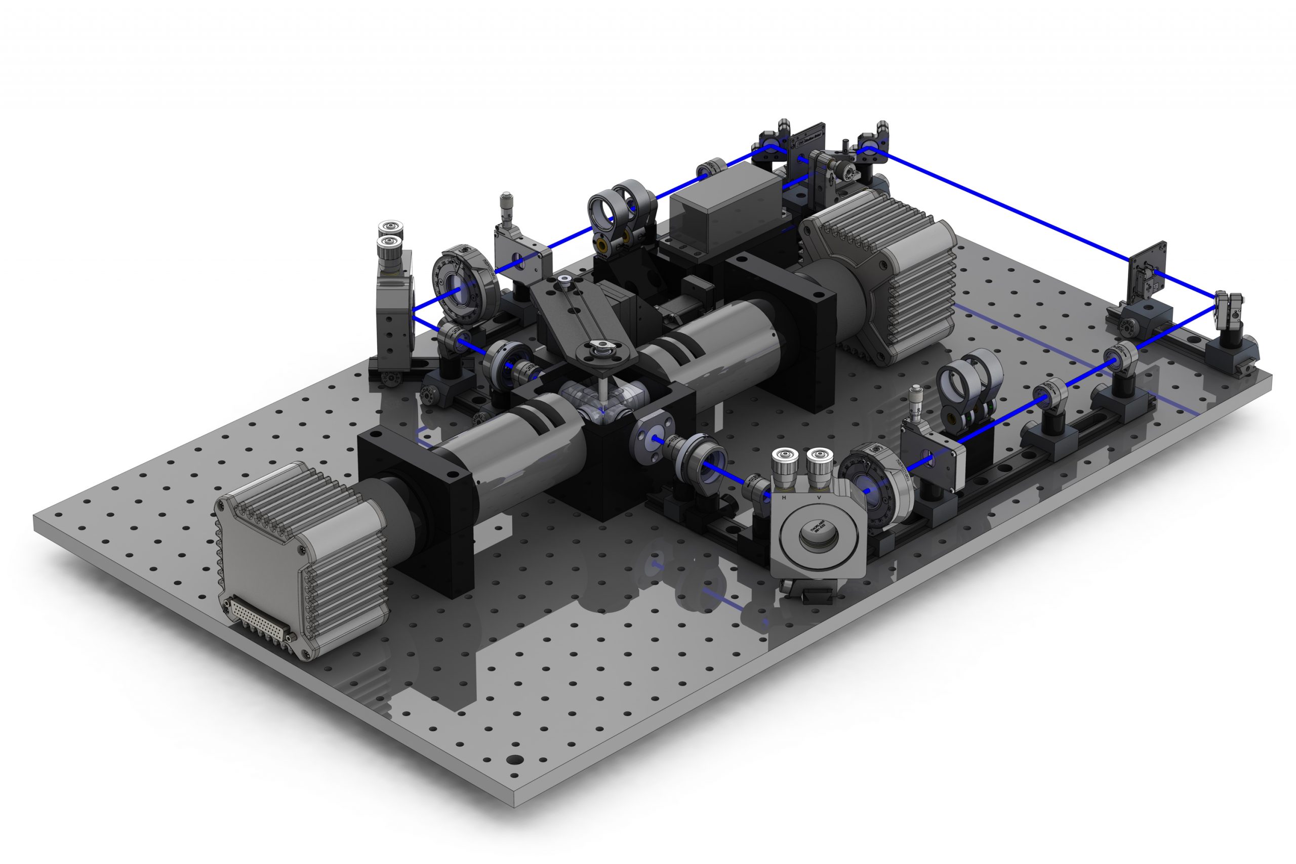
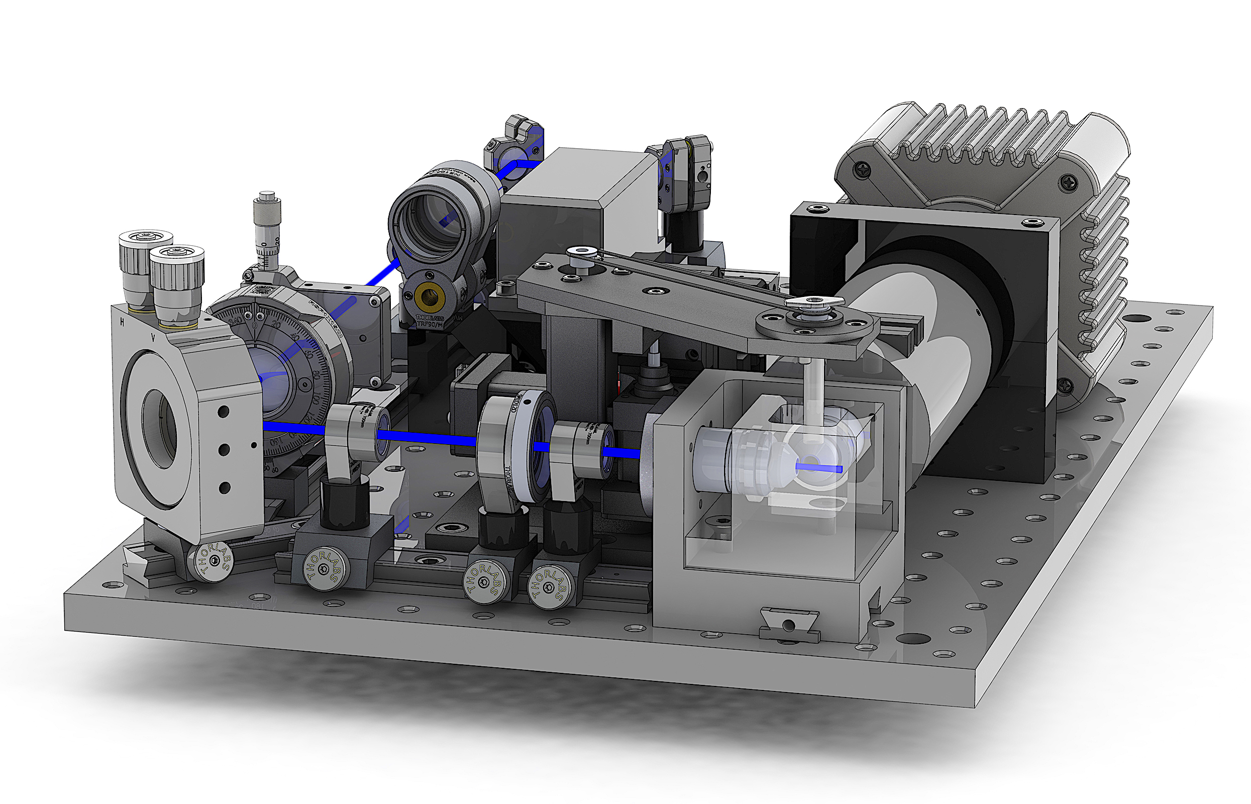
Lightsheet fluorescence microscopy (LSFM) is a type of fluorescence microscopy that uses a thin sheet of laser light to excite fluorescent molecules within a biological specimen. LSFM allows for high-resolution 3D imaging of living cells and tissues, with minimal phototoxicity and photobleaching compared to other fluorescence microscopy techniques.
In LSFM, the laser light is directed perpendicular to the imaging axis, illuminating only a thin section of the specimen at a time. The emitted fluorescence is then captured by a high-resolution camera, producing a 3D image of the specimen. By imaging the specimen from different angles and combining the images, a full 3D reconstruction of the specimen can be created.
LSFM has numerous applications in biological research, including developmental biology, neuroscience, and cancer research. It allows for the study of complex biological processes in living organisms, such as the formation of organs and tissues, the growth and movement of cells, and the interactions between cells and their environment.
Overall, LSFM is a powerful imaging technique that has revolutionized the field of biology and has the potential to significantly advance our understanding of complex biological systems.