High-Resolution & High-Power Fluorescence In Vivo Imaging-10523

Imaging Modes: Brightfield, Drakfield, Polarizing, Fluorescence, Phase Contrast, DIC Nomarski

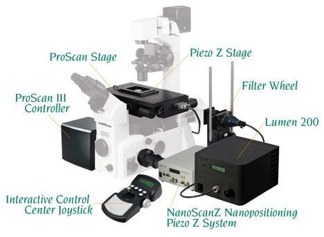
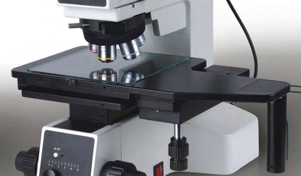
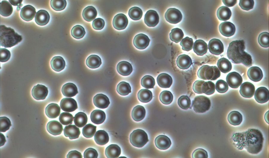
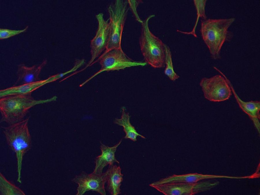
Microscopes are used today by people from mid / high school students to the scientists and in several fields. It is even used as a daily routine work by biologists, physicians / hospital laboratory, geologists, dentists / dental technicians, veterinarians, paleontologists, entomologists, gemologists, hair transplant, scientific researchers, quality control personnel, quality assurance, assemblers, forensic document […]

BIO-WCAM Series C-mount WIFI CMOS Camera-10470

Super Fast 1 Billion/Trillion FPS Ultra High-Speed HD Camera

1.000.000.000.000 (1 Billion/Trillion) FPS!!! Ultra High-Speed Camera HD 1.000.000.000.000 (1 Billion/Trillion) FPS!!! Ultra High-Speed Camera HD 1000000000000 (1 Billion/Trillion) FPS!!! Ultra High-Speed Camera (Licht/Light) HD 720p 1 Frame = 1.71 picoseconds Researchers at MIT have created a camera with a resolution of one frame per trillionth of a second, allowing them to film light as […]

Polarizing Microscopy Imaging (Basics & Applications)

Brightfield Image: Brightfield (BF) is the simplest imaging technique of all the optical microscopy imaging mode. As the name states you can expect an image with bright background. A simple example in our daily life is like seeing birds or airplane in the sky. In BF imaging, a sample is normally illuminated via transmitted light […]

Fluorescence Microscopy

Fluorescence Microscopy The fluorescence microscopy developed to one of the most important tools of the life science area. The observation of the fluorescence emergences in biological samples delivers differentiates conclusions about morphology and functionality of cells. The objects of examination reach from fixated cells via chromosomes and genes up to observation of processes in living […]

Live Cell Imaging

Live Cell Imaging Live cell imaging is the study of the life of living cells / organisms using a time-lapse video microscopy in which the cells are kept at the closest conditions to a regular laboratory incubator. Live cell imaging is used by scientists and researchers to obtain a better understanding of dynamic events occur […]

Metallurgical Microscopy Basics & Applications

What is a metallurgical microscope? A simple answer is that a metallurgical microscope is another type of light microscopes and unlike a biological microscope it uses a reflected white light. Obviously the nature of samples is different for such a use, i.e. a metal, semiconductor or plastic rather than a biology slide, cell, living microorganism […]

Phase Contrast Microscopy

What is Phase Contrast Microscopy? Phase Contrast is a contrast enhancing optical microscopy technique in which an unstained transparent sample is seen with high brightness by shifting phase difference of light. The idea of Phase Contrast Imaging was brought by Frits Zernike in 1930’s, a Dutch physicist, who got the Nobel prize in 1953 for […]

Sample Microscope Images

Captured using our IncuScope with 3 LEDs (UV, Blue and Green), 3MP sCMOS camera BIC-C30, and 20x objective lenses, and IncuView software. Sample: FluoCells® Prepared Slide #2: bovine pulmonary artery endothelial cells (BPAE) cells with Mouse Anti-α-tubulin, BODIPY FL Goat Anti-Mouse IgG, Texas Red-X Phalloidin, and DAPI), (Molecular Probes) Further details: https://www.bioimager.com/product/oem-microscope-inverted-upright-for-biological-industrial-application/

Microscope History

The oldest history of microscope goes to 1590s, when two Dutch spectacle makers, Zacharias Jansen and his father Hans Jansen, started some tests with lenses. They combined several lenses and put inside a tube and boom, they made the discovery. They saw an object at the end of the tube can bee seen way larger […]

Fast Worldwide shipping

Your order will be shipped ASAP

Easy 10 days returns

10 days money back guarantee

International Warranty

Offered in the country of usage

100% Secure Checkout

PayPal / MasterCard / Visa