Microscopy Imaging of a Virus
These days there are lots of discussions on virus, particularly on novel coronavirus COVID-19. Scientists and ordinary people like to know the image of this, as so many calls it enemy. Here we review the possible methods to see the available images of viruses in general and COVID-19 in particular using microscopy method.
Introduction to Virus
Viruses are one of the smallest living microorganisms. They cannot grow or proliferate on its own, thus often called particles, but they can survive for hours, days, months and even years at some conditions (like frozen temperature and dry) and act as an infectious agent. History of virus science goes to 1892 when Dmitri Iosifovich Ivanovsky (Russian Scientist, 1864-1920) described it as a non-bacterial pathogen that infected tobacco plants. Today after 128 years, over 5,000 virus species have been classified and described in the scientific journals. The origins of viruses are not clear yet. Some developmental biologists, evolutionist and virologists think it may have evolved from plasmids (a piece of DNA that can move between cells) or developed from bacteria. Viruses have life because they carry genetic materials, can reproduce and develop although they do not have cell structures to be counted as life, i.e. described as “organisms at the edge of life”.
Due to infectious and replication capability of viruses, electronic engineers and computer experts use “computer virus” term that infects the digital systems, personal computers, smartphones and networks, not discussed here. Interestingly, one of the scientists was able to capture its image (TBD).
Regardless of type, both living viruses and computer viruses cause millions and billions of dollars worth of economic damage each year.
Size
Bacterium (singular form of bacteria) is another unicellular organism, which can be seen under a light microscope. Bacteria has a size of one-tenth (1/10) of a living cell which is 0.5 to 5.0 micrometers (µm) in length (its shape can be cylindrical, spherical,…). Viruses have a big diversity of shapes and sizes, i.e. morphogenesis. Viruses are way smaller than bacteria and have size range of one-tenth and even one-hundredth of most bacteria. The most studied viruses have a diameter range of 20 to 350 nanometers (nm) or 0.020-0.350µm. There are some exceptions, for instance filoviruses have a length of up to 1400nm or 1.4um and diameter of 80nm / 0.080µm. Mimivirus is one of the largest size and has 390nm / 0.39µm capsid diameter.
Visualization of a virus
Due to their smaller size, most of viruses cannot be seen using a light microscope. However, a giant type of viruses such as Megavirus chilensis, which has dsDNA, was seen with a basic light microscope in 2011 by Defne Arsalan et al.
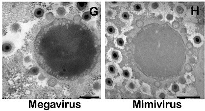 Figure 1. Electron microscopy of Megavirus and Mimivirus. Scale bars: 1μm. Reference: Arsalan et al. PNAS, 2011, 108, 42, 17486-91.
To visualize the viruses, some of the techniques have been suggested in the literature.
1. Light Microscopes
A standard light microscope has 1000x total magnification, which uses a 100x objective lens and 10x eyepieces. There are some ways to go beyond this number. For instance, Bioimager has a pending patent that shows how to reach over 5000x magnification in a typical microscope. This includes combination of a high-magnification objective lens with the second high-magnification lens, which is a C-mount or T-Mount adapter, connecting to a digital camera. The total magnification can be from 5,000x till 10,000x. This requires a high-power illumination, high sensitive camera and super fine focus mechanism. The problem is the resolution limit forced by the wavelength of a visible light (400-700nm). The image can be enlarged but the resolution can luckily be at the wavelength size, i.e. 200nm or 0.2µm.
2. Fluorescence Microscopy and Confocal Microscope
This method uses fluorphores or fluorochromes which allows organisms to luminate fluorescently when a proper light shines and suitable optics are combined. This method is mainly used to track virally infected DNA in a host cell. This means we observe a population of virus. This method can help for quantitative estimation of viral concentration in a given sample.
3. Total Internal Reflection Dark-Field Microscopy (TIRDFM)
This is a special optical imaging technique and utilizes light scattering from an evanescent field. Its advantages are generating a high-spatial resolution and high signal-to-noise ratio in the vertical dimension. It is highly used for particle tracking. This technique is label-free but often fluorophors are introduced into the sample for better and much more selective visualization. In that case it is called Total Internal Reflection Fluorescence Microscopy (TIRFM).
Anyhow, this microscopy system can be achieved by using a perforated mirror in place of a dichroic mirror. Sample preparation follows a relatively complicated protocol. When it comes to observation, you can see blight spots with a dark background.
4. Transmission Electron Microscopy
A Transmission Electron Microscope (TEM) allows 1000 times better resolution than an ordinary light microscope. Thus, viruses can be easily observed. A higher resolution is achieved with TEM because it utilizes an electron beam with 0.005nm diameter instead of a large wavelength light in optical microscopy. The problem is preparation of the sample which needs carefully processed. When it comes to a final image, viruses can be easily seen like particles inside a cell (where they infected the cell).
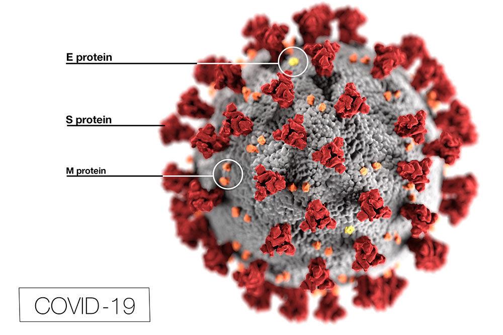
Coronavirus image captured by an electron microscope; illustration credit: the Centers for Disease Control (CDC).
Recent Findings
Russian scientists at the Vector Institute in Novosibirsk were able to take the microscope images of the new coronavirus COVID-19 on Thu Mar 19, 2020. A sample taken from an infected patient by specialists of the Russian Influenza Research Institute and a microscopist in Rospotrebnadzor captured the image using negative contrast method on a transmission electron microscope JEM-1400. They stated that the size of the particle was 100-120nm / 0.1-0.12um.
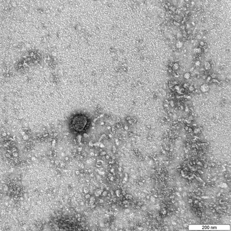
Video: https://www.youtube.com/watch?v=mGdDuI0BZOQ
Using scanning and transmission electron microscopy, a team in NIAID (The National Institute of Allergy and Infectious Diseases), USA captured the image of novel corona virus COVID-19 from a patient in US. The microscopist Elizabeth Fischer generated the image and published in Feb 13, 2020.
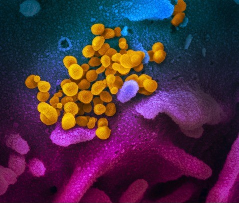 Scanning electron microscope image of Novel coronavirus COVID-19 (yellow) emerging from the surface of cells (blue/pink) cultured in the lab. Credit: NIAID-RML Feb 13, 2020.